
Leukoplakia is a white patch in the mouth that cannot be wiped away or explained by another condition, and erythroplakia is a velvety red patch of the same kind. Both are classed as potentially malignant disorders, which means they are not cancer themselves but carry a higher-than-normal chance of changing over time. Any patch that has not cleared within two weeks deserves a professional check, because catching change early is what keeps treatment simple.

Key takeaways
- Leukoplakia is a persistent white patch, erythroplakia is a persistent red patch, and neither wipes off.
- Both are potentially malignant disorders, so they are watched and often biopsied.
- Red patches (erythroplakia) carry a much higher risk than white patches and need prompt assessment.
- Smoking, alcohol, and a history of either greatly raise the risk, and stopping smoking can help patches resolve.
The single rule: a patch that lasts more than two weeks gets checked
The most important habit is simple. If you find a white or red patch in your mouth that has not gone within two weeks, have it examined. Most patches turn out to be harmless, such as a friction line from a sharp tooth or a denture rub. But because a small number are an early warning, the safe approach is to let a dentist decide rather than wait and watch at home.
Leukoplakia: the white patch
Leukoplakia appears as a white or greyish patch that cannot be scraped off and is not caused by an obvious irritation. It is often painless, which is why it can go unnoticed. Patches that are flat and even (homogeneous) carry a lower risk, while patches that are lumpy, mixed red and white, or wart-like (non-homogeneous) carry a higher risk. Research puts the overall long-term rate of change to cancer at roughly 7 percent, with figures around 3 percent for the flat even type and up to about 14 percent for the mixed or lumpy types.
Erythroplakia: the red patch
Erythroplakia is less common but far more concerning. It appears as a smooth, velvety red patch, often on the floor of the mouth, the underside of the tongue, or the soft palate. Studies report high rates of change, with estimates ranging widely and an average that can be considered around 30 percent. Importantly, a large share of red patches already show significant change at the first biopsy, which is exactly why a red patch should never be left to see if it settles.
| Feature | Leukoplakia | Erythroplakia |
|---|---|---|
| Colour | White or grey | Red, velvety |
| Wipes off? | No | No |
| Pain | Usually none | Usually none |
| Relative risk of change | Lower (around 7 percent overall) | High (often around 30 percent) |
| Action | Assess, often biopsy | Prompt biopsy |
What raises the risk
The strongest risk factors are smoking and heavy alcohol use, and the two together multiply the risk. Chewing tobacco and betel quid are also strongly linked. Our articles on smoking and oral health and vaping and oral health explain how these habits affect the mouth lining over time. Stopping smoking is one of the few changes that can help some patches shrink or disappear.
How they are assessed
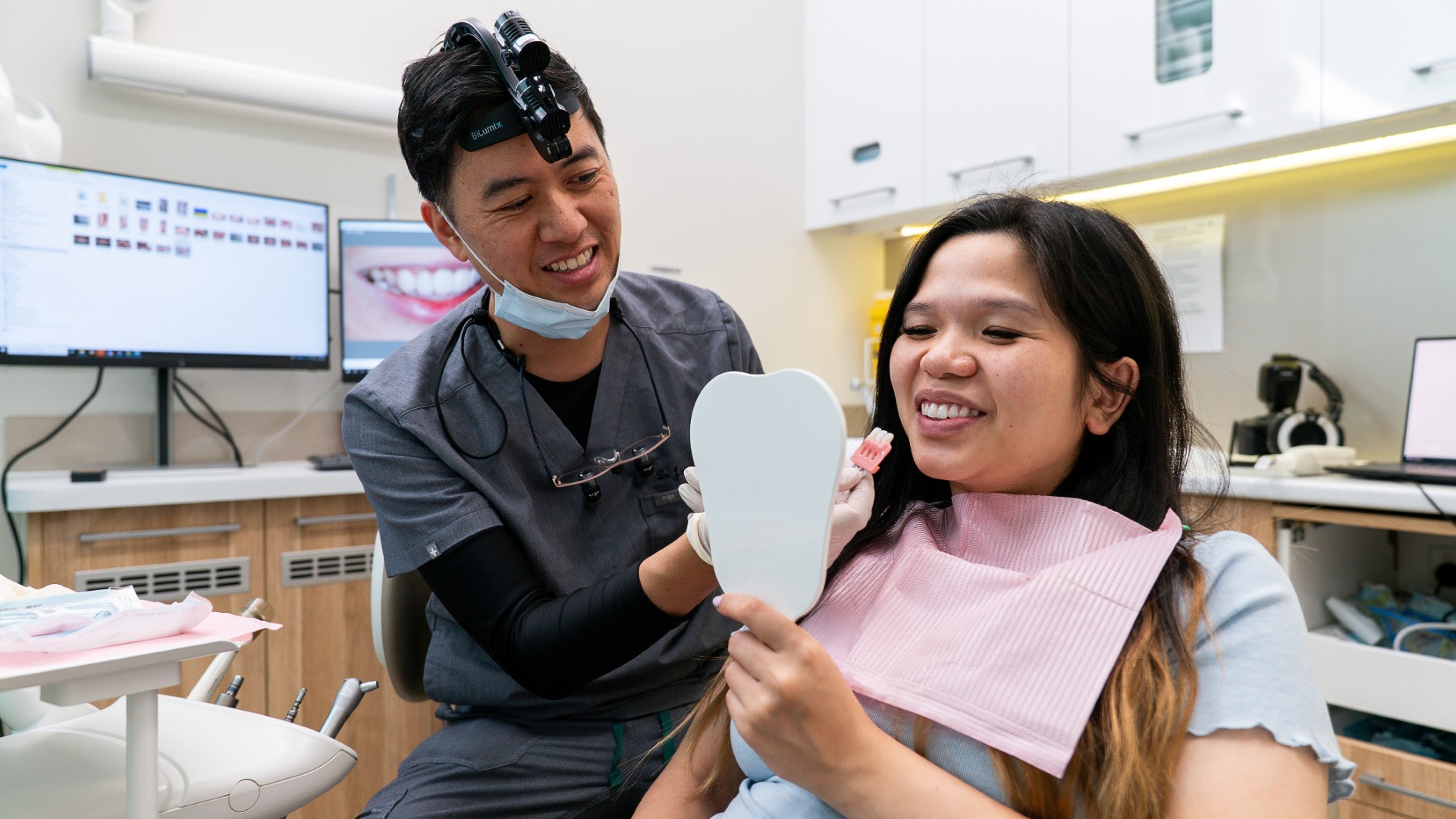
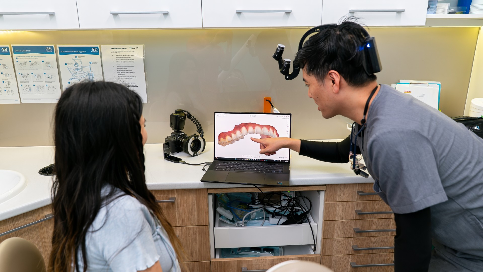
A dentist examines the patch, notes its colour, texture, and site, and removes anything that might be irritating it, such as a sharp filling edge or a rough denture. If a white patch persists after the irritant is removed, or if there is any red component, a biopsy is taken to look at the cells. This is the only way to know for sure whether the patch is harmless, showing early change, or needs more action. This careful checking is part of routine oral cancer screening, which we recommend at regular check-ups, especially for people who smoke or drink.
How they are managed
Management depends on the biopsy result. Low-risk patches may simply be monitored with photographs and regular reviews. Higher-risk patches are often removed, by surgery or laser, and then watched, because even removed lesions can return. Alongside this, reducing risk factors matters, so support to stop smoking and cut down alcohol is part of good care. Patches can recur, so ongoing review is normal rather than a sign that something went wrong.
General cost and what to expect
Assessment usually involves an examination and a biopsy, with any removal and follow-up depending on the result. Because fees vary with what is needed, we do not list our own prices here. You are welcome to view current options on our deals and pricing page or arrange a written quote at a general dental consultation.
Frequently asked questions
Are leukoplakia and erythroplakia cancer?
No. They are potentially malignant disorders, meaning they carry a raised risk of change but are not cancer themselves. Most do not become cancer, which is why monitoring and prompt biopsy matter.
Which is more serious, a white patch or a red patch?
A red patch (erythroplakia) is more concerning. It is less common but carries a much higher risk of change, so it should be checked promptly.
Can leukoplakia go away on its own?
Some patches improve after an irritant is removed or after stopping smoking. Others persist and need treatment. A dentist can tell you which is likely after examining it.
How is this different from oral thrush?
Thrush is a fungal infection and the white coating usually wipes off, leaving a red base, and it responds to antifungal treatment. Leukoplakia does not wipe off. A dentist can tell them apart.
Do I need a biopsy?
Often yes, particularly for any red patch or a white patch that persists. A biopsy gives a definite answer rather than guessing from appearance alone.
When to see a dentist
See a dentist promptly for any patch, lump, or sore in the mouth that lasts more than two weeks, especially a red patch or one that is mixed red and white. The team at Lumi Dental can examine it, arrange a biopsy, and guide the next steps. Book through our contact page.
This article is general information and is not a substitute for individual advice. Persistent mouth changes should always be assessed by a dentist or doctor.