

Dens invaginatus, sometimes called a tooth within a tooth, is a developmental fold where the surface of a tooth folds inward before it erupts, creating a pocket inside the tooth that can trap bacteria and lead to early infection. On an X-ray this fold can look like a small tooth sitting inside the main one, which is where the name comes from. It most often affects the upper lateral incisors, the teeth just to the side of the two front teeth. The importance is practical: the fold can give bacteria a direct path to the nerve, so finding and sealing it early can protect the tooth.
Key takeaways
- Dens invaginatus is an inward fold formed as a tooth develops.
- It looks like a tooth within a tooth on an X-ray.
- It most often affects the upper lateral incisors.
- The fold can trap bacteria and lead to early nerve infection.
- Early sealing or treatment can protect the tooth.
What it is
As a tooth forms, the outer enamel layer normally shapes a smooth crown. In dens invaginatus, part of that surface folds inward before the tooth is complete, leaving a pocket lined by enamel that extends into the tooth. Because the pocket connects to the outside, food and bacteria can collect in it. In more pronounced cases the fold reaches deep toward or into the nerve space, which is why the condition matters more than its harmless appearance suggests. The tooth usually looks normal or only slightly unusual from the outside, so it is often found on an X-ray.

The three types
Dentists group dens invaginatus into three types based on how deep the fold goes, using a system described by Oehlers. The type guides how closely a tooth needs watching and how it is treated.
| Type | How deep the fold goes | What it means |
|---|---|---|
| Type 1 | Within the crown only | Mildest and most common, easier to seal |
| Type 2 | Extends into the root canal area | May connect with the nerve, closer monitoring |
| Type 3 | Passes through and opens near the root tip | Most complex, higher infection risk |
Type 1 is the most frequently seen. The deeper types carry a higher risk of the nerve becoming infected, sometimes even before decay is visible, which is why early diagnosis is valuable.
Why it matters
The main concern is that the fold gives bacteria a sheltered route toward the nerve. A tooth with dens invaginatus can develop infection and an abscess at a young age, sometimes while the tooth still looks healthy on the surface. Catching it early means a dentist can seal the vulnerable pit with a filling before bacteria get in, much like sealing the deep grooves of a molar. If the nerve is already affected, root canal treatment may be needed, and the unusual internal shape makes that work more complex, so planning with X-rays and sometimes 3D imaging helps.
How it is found and managed
Dens invaginatus is usually discovered on a routine X-ray or when a young tooth develops unexpected infection. Once identified, management depends on the type and whether the nerve is involved. A mild fold with a healthy nerve can often be sealed early to keep bacteria out. Where infection has reached the nerve, root canal treatment or, in complex cases, a minor surgical procedure at the root tip may be needed to save the tooth. Very occasionally a tooth cannot be saved and is replaced, and our guide to replacing a missing tooth explains the options. Good prevention throughout, covered in our guide to preventing decay, protects the tooth, and it sits alongside other developmental variations like taurodontism.
Frequently asked questions
What is dens invaginatus?
It is a developmental fold where a tooth surface folds inward before eruption, creating a pocket inside the tooth. On an X-ray it can look like a tooth within a tooth.
Which teeth are usually affected?
The upper lateral incisors, beside the two front teeth, are most often affected, though other teeth can be involved.
Why is it a problem?
The fold can trap bacteria and give them a path toward the nerve, so an affected tooth can become infected early, sometimes while it still looks healthy.
How is it treated?
A mild case can be sealed early to keep bacteria out. If the nerve is infected, root canal treatment or minor surgery may be needed, guided by X-rays.
Can the tooth be saved?
Usually yes, especially when found early. Sealing or timely root canal treatment saves most teeth, though complex cases need careful planning.
Speak with the team at Lumi Dental
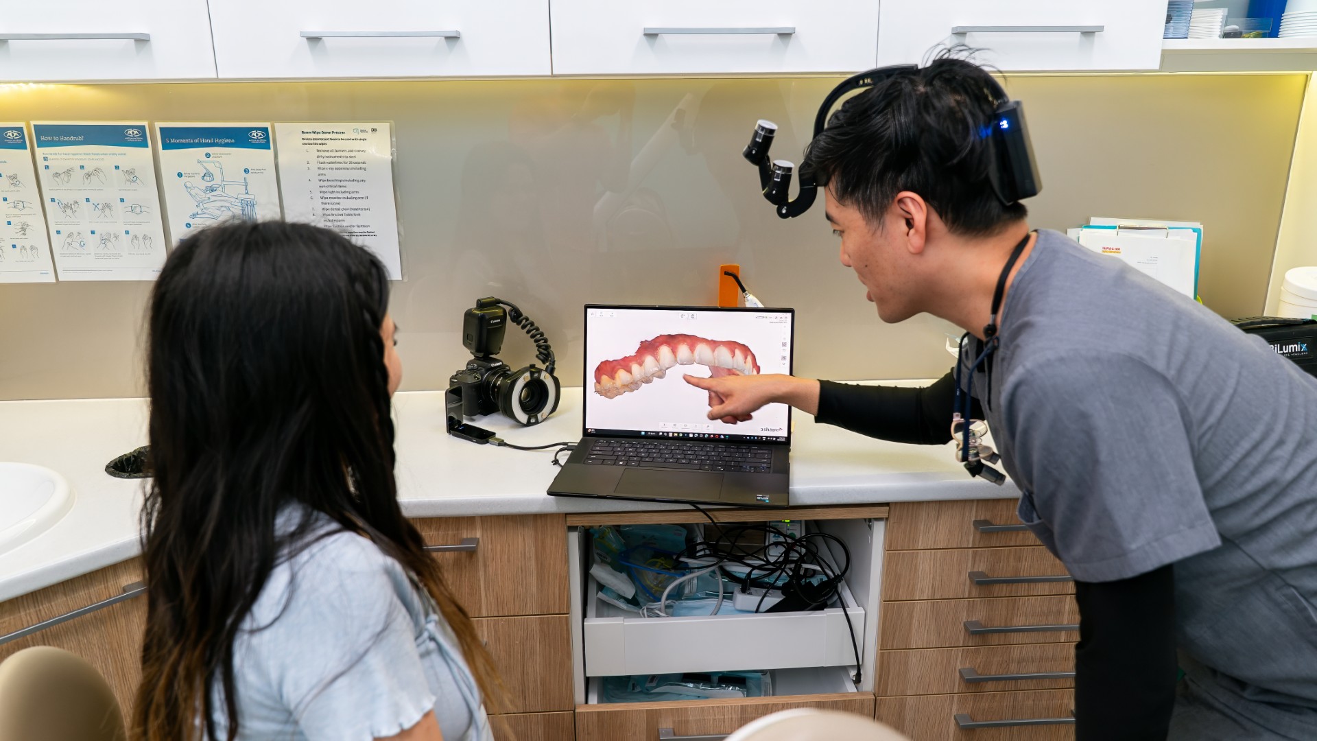
If an X-ray has shown a tooth within a tooth, the team at Lumi Dental in Melrose Park can explain what it means and plan any care needed. Read more about general dental care or view current new-patient offers on the current deals page. We do not list our own prices here and are happy to provide a written quote after an assessment.
This article is general information only and is not a substitute for personal dental advice. Please see your dentist for advice about your situation.