A pink patch showing through a tooth near the gum is usually not a surface stain, and brushing or whitening will not remove it. It can be a sign that tooth structure is being lost and replaced by soft tissue, a process dentists call resorption. Because it is often painless, it is easy to put off, but the size of the defect when it is found has a large bearing on which treatments are possible.
The colour is tissue showing through the tooth
Resorption is the loss of dental hard tissue through the action of odontoclasts, cells that break down mineralised tissue. When the resorbing area comes close to the surface of the crown, the tissue inside it, which has a rich blood supply, can show through the enamel as a pinkish shade.
Dentists have long called this the "pink tooth of Mummery" and linked it with resorption that starts inside the tooth. A 2026 review in the Journal of Clinical and Experimental Dentistry (Mercante and colleagues) notes that the pink colour is not exclusive to internal resorption and can also occur when the process starts on the outside of the root. So the colour alone does not show where the process began.
Two kinds of resorption that start in different places
The distinction matters because the two forms are usually treated differently.
| Internal resorption | External cervical resorption | |
|---|---|---|
| Where it starts | Inside the root canal | On the root surface just below the gum attachment |
| The nerve of the tooth | The process starts in the pulp, which needs to be at least partly alive for it to continue | Often still alive; a Taiwanese study of 63 affected teeth found 73% had a vital pulp |
| Most often linked with | Past injury to the tooth | Braces, injury and bleaching inside a root-filled tooth, alone or combined |
| Usual first treatment | Root canal treatment in suitable cases | Depends on the extent; often a repair from the outside, sometimes with root canal treatment |
Internal resorption is described in the Mercante review, which found trauma the most frequently reported factor and conventional root canal treatment the main approach in favourable cases. The rest of this article focuses on external cervical resorption, which can also show as a pink spot near the gum line.
External cervical resorption often causes no symptoms
External cervical resorption begins on the root surface just under the gum. In a study of 27 affected teeth that were still alive, Mavridou and colleagues (Journal of Endodontics, 2016) found that it starts in the cementum below the gum attachment and spreads into the tooth in three dimensions. In these teeth a thin resistant layer around the nerve kept the resorption from breaking into the pulp, which may be one reason many affected teeth do not hurt.
That fits what clinics report. In a Taiwanese series of 63 affected teeth (Jeng and colleagues, Journal of Endodontics, 2020), most teeth had no pain on tapping or pressing, no deep gum pocket and no abscess. The authors concluded that affected teeth typically lack signs and symptoms and that X-rays are critical for early diagnosis. Deep pockets and abscesses were seen in advanced cases.
Upper front teeth are affected more than others. In a Belgian and Dutch series of 337 teeth (Mavridou and colleagues, Journal of Endodontics, 2017), 29% were upper central incisors and 72% were upper teeth.
It is still uncommon. Across three private endodontic practices in the United States (Huang and colleagues, Clinical Oral Investigations, 2023), it was found in under 1% of patients seeking root canal care, although the rate more than doubled, from 0.46% in 2010 to 2015 to 0.99% in 2016 to 2021. A later paper by Patel and colleagues notes that wider use of 3D X-rays has been suggested as one reason more cases are being found.
Braces, knocks and internal bleaching are the factors studies keep finding
The cause is not fully understood, and studies report associations rather than proof. The same few factors appear again and again:
- Orthodontic treatment. In a study by Heithersay at the University of Adelaide (Quintessence International, 1999), orthodontics was the most common single factor, present alone in 24.1% of 257 affected teeth.
- Dental injury. Trauma alone was the second most common factor in that study (15.1% of teeth). A 2026 systematic review in Dental Traumatology (Lima and colleagues) found trauma reported in 8.5% to 33.3% of cases across eight studies, but concluded the data cannot establish cause.
- Bleaching inside a root-filled tooth. Heithersay found intracoronal bleaching as the only factor in 3.9% of teeth, and combined with trauma or braces in a further 9.7%.
In many cases more than one factor is present. Mavridou and colleagues found more than one in 59% of teeth, while in Heithersay's group 16.4% of teeth had no identifiable factor at all. A 2025 London study of 215 affected teeth (Patel and colleagues, International Endodontic Journal) found no identifiable factor in 22.3% and also recorded cat ownership in 15.8%. Cats commonly develop a resorptive tooth condition of their own, and a link through a cat virus has been suggested, but it has not been tested or shown.
If you have had braces or an injury, this is not usually a reason for concern, since the condition is uncommon. It is a reason to keep up regular check-ups, especially for a tooth that was knocked. Our articles on root resorption after braces (a different type that shortens root tips) and on teeth replanted after an injury cover those situations. If a dark tooth has been whitened from the inside, our page on internal bleaching explains that treatment.
X-rays, sometimes in 3D, confirm what is going on
A dentist will usually look at the spot, gently probe the gum around it and take an X-ray. Taking two X-rays from slightly different angles can help separate the two types: a defect that starts outside the root appears to shift when the angle changes, while one inside the canal stays centred on it.
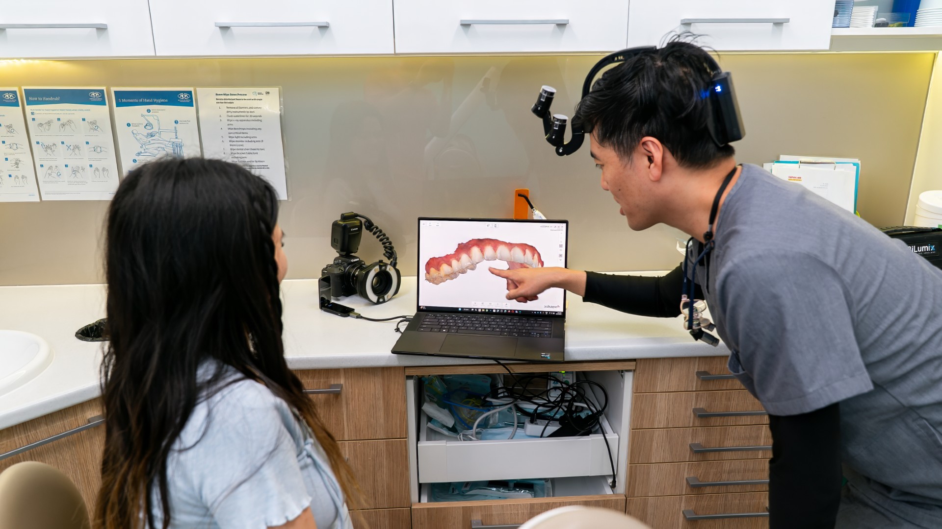
A standard X-ray is flat, so it can miss how far the defect has spread around the root. A 2026 meta-analysis in the Australian Endodontic Journal (de Sousa and colleagues) found that cone-beam CT, a 3D dental scan, detected external cervical resorption more accurately than standard X-rays, although the certainty of that evidence was rated very low because only four studies qualified. Lumi Dental has an Orthophos 3D X-ray unit, so a 3D X-ray can be taken in the practice when it is needed; our technology page explains how these scans are used.
Treatment depends on how far it has spread
Heithersay grouped these defects into four classes, from a shallow defect near the gum (class 1) to one that has spread beyond the upper third of the root (class 4). In his treatment study of 101 teeth followed for at least three years, all class 1 and class 2 cases were judged successful, 77.8% of class 3 cases, and only 12.5% of class 4 cases. A 2026 narrative review from Adelaide (Rossi-Fedele, Doğramacı and Heithersay, International Endodontic Journal) found that across six later studies, classes 1 and 2 were linked with high survival and class 4 with the opposite.
A review of management by Patel and colleagues (International Endodontic Journal, 2018) describes the usual options:
- A small defect above the bone can often be cleaned out and sealed from the outside, without root canal treatment.
- A larger defect may also need treatment of the nerve, or a repair from inside the tooth with root canal treatment when the outside cannot be reached.
- In selected cases where neither approach is practical, the tooth may be removed, repaired and replaced in its socket.
- Where treatment is not practical, the tooth may be monitored, and an untreatable tooth may need to be removed.
Some published case reports describe repairs with bioceramic materials. Lumi uses bioceramic materials, including Biodentine, for pulp protection and root repair. Depending on how far the defect reaches, your dentist may also recommend referral to an endodontist, a registered specialist in root canal and root problems.
Results over many years vary. In a series of 313 teeth that were treated or monitored (Mavridou and colleagues, International Endodontic Journal, 2022), about 70% were still present at five years, survival fell with longer follow-up (about 29% at ten years), and outcomes depended on how extensive the defect was. If a tooth cannot be kept, a dental implant is one of the ways to replace it.
Questions about pink spots and resorption
Is a pink spot an emergency?
It often causes no pain, so it does not usually need same-day care. It does need a prompt examination, because a smaller defect is generally easier to repair than a larger one. Swelling, an abscess or pain are reasons to be seen sooner.
Will whitening hide it?
No. The colour comes from tissue inside the defect, not from a surface stain, so whitening gel will not change it. A tooth with resorption should be assessed before any whitening.
Does it spread to other teeth?
In the Taiwanese series, most patients had a single affected tooth, although the 31 patients had 63 affected teeth between them, so some had several. Other teeth with the same history, such as several that were moved with braces or injured together, may be checked on the X-rays at the same visit.
Having a pink spot checked
If you have noticed a pink area near the gum on one of your teeth, book an examination with a general dentist at Lumi Dental in Melrose Park. Your dentist can examine the tooth, take any X-rays needed and explain the options. You can book online or contact the practice.
Sources
- Patel S, Kanagasingam S, Pitt Ford T. External cervical resorption: a review. J Endod. 2009;35(5):616-25. PubMed 19410071
- Mercante HS and colleagues. Internal root resorption: understanding the pink tooth of Mummery and its clinical implications. J Clin Exp Dent. 2026;18(5):e616-e627. PubMed 42434214
- Mavridou AM and colleagues. Understanding external cervical resorption in vital teeth. J Endod. 2016;42(12):1737-1751. PubMed 27776885
- Jeng PY and colleagues. Invasive cervical resorption: distribution, potential predisposing factors, and clinical characteristics. J Endod. 2020;46(4):475-482. PubMed 32115249
- Mavridou AM, Bergmans L, Barendregt D, Lambrechts P. Descriptive analysis of factors associated with external cervical resorption. J Endod. 2017;43(10):1602-1610. PubMed 28807370
- Huang J and colleagues. The prevalence, characteristics, and risk factors of external cervical resorption: a retrospective practice-based study. Clin Oral Investig. 2023;27(9):5595-5604. PubMed 37493728
- Heithersay GS. Invasive cervical resorption: an analysis of potential predisposing factors. Quintessence Int. 1999;30(2):83-95. PubMed 10356560
- Heithersay GS. Treatment of invasive cervical resorption: an analysis of results using topical application of trichloracetic acid, curettage, and restoration. Quintessence Int. 1999;30(2):96-110. PubMed 10356561
- Lima TFR and colleagues. Traumatic dental injuries as a potential predisposing factor for invasive cervical resorption: a systematic review. Dent Traumatol. 2026 (online ahead of print). PubMed 42885097
- Patel S and colleagues. Potential predisposing features of external cervical resorption: an observational study. Int Endod J. 2025;58(2):273-283. PubMed 39560698
- de Sousa SEM and colleagues. Diagnostic accuracy of cone-beam computed tomography and periapical radiography in detecting external cervical resorption: a systematic review and meta-analysis. Aust Endod J. 2026;52(1):272-281. PubMed 41273117
- Rossi-Fedele G, Doğramacı EJ, Heithersay GS. Prognostication of external cervical resorption based on Heithersay's and Patel et al. classifications: a narrative review. Int Endod J. 2026;59(7):1376-1383. PubMed 41689254
- Patel S, Foschi F, Condon R, Pimentel T, Bhuva B. External cervical resorption: part 2, management. Int Endod J. 2018;51(11):1224-1238. PubMed 29737544
- Mavridou AM and colleagues. A clinical approach strategy for the diagnosis, treatment and evaluation of external cervical resorption. Int Endod J. 2022;55(4):347-373. PubMed 35034370
- Ghabraei S, Bolhari B, Hashemi N, Gharehchahi H. Conservative management of an advanced external cervical resorption with internal approach using bio-ceramic materials: a case report. Clin Case Rep. 2024;12(1):e8378. PubMed 38161643
- Kumari V and colleagues. Conservative management of invasive cervical resorption using Biodentine with adjunctive laser-assisted photodynamic and photobiomodulation therapy: a case report. Cureus. 2026;18(4):e107364. PubMed 42170158
This article is general information only and is not a substitute for advice about your own situation from a dentist.