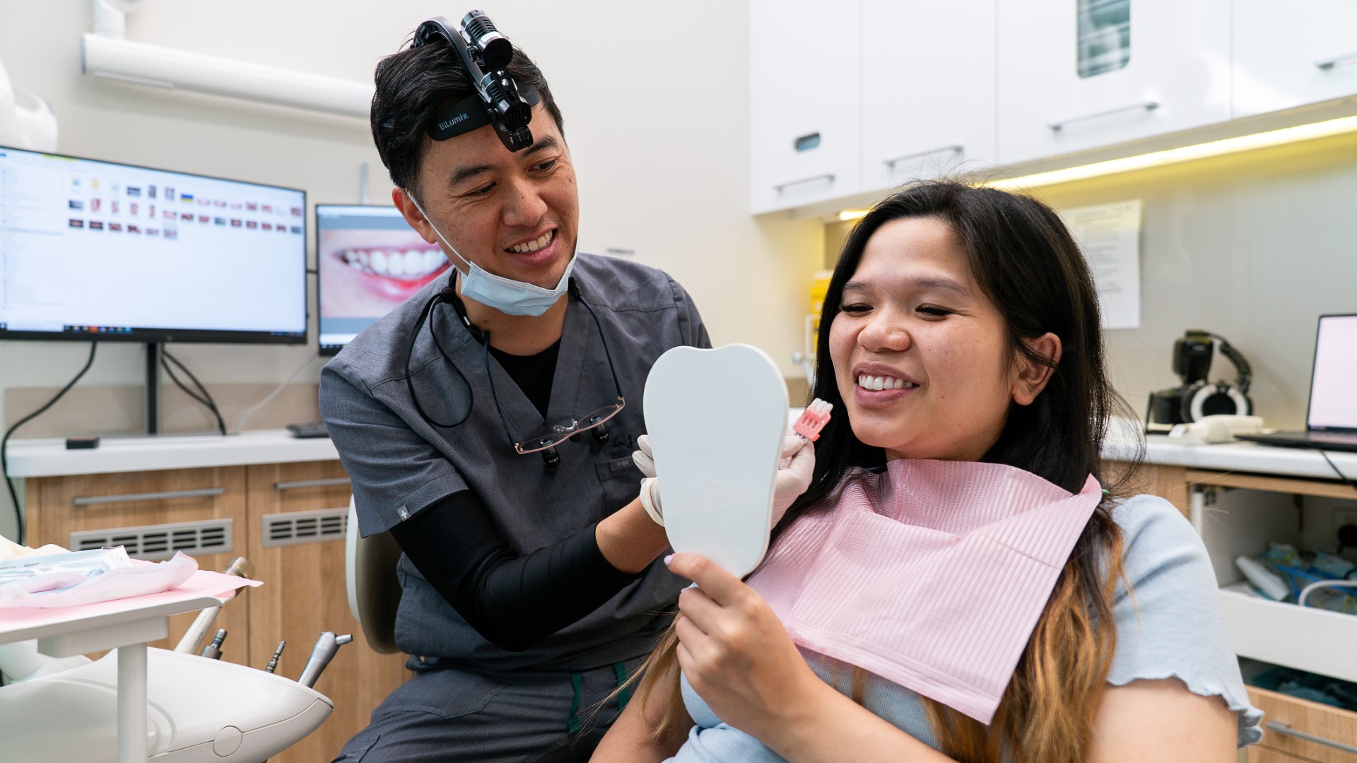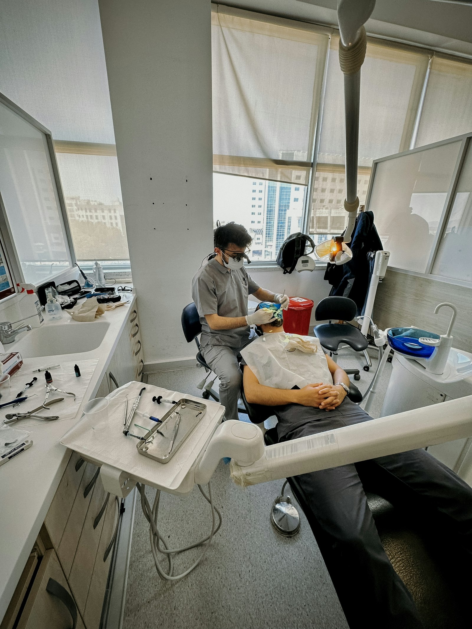
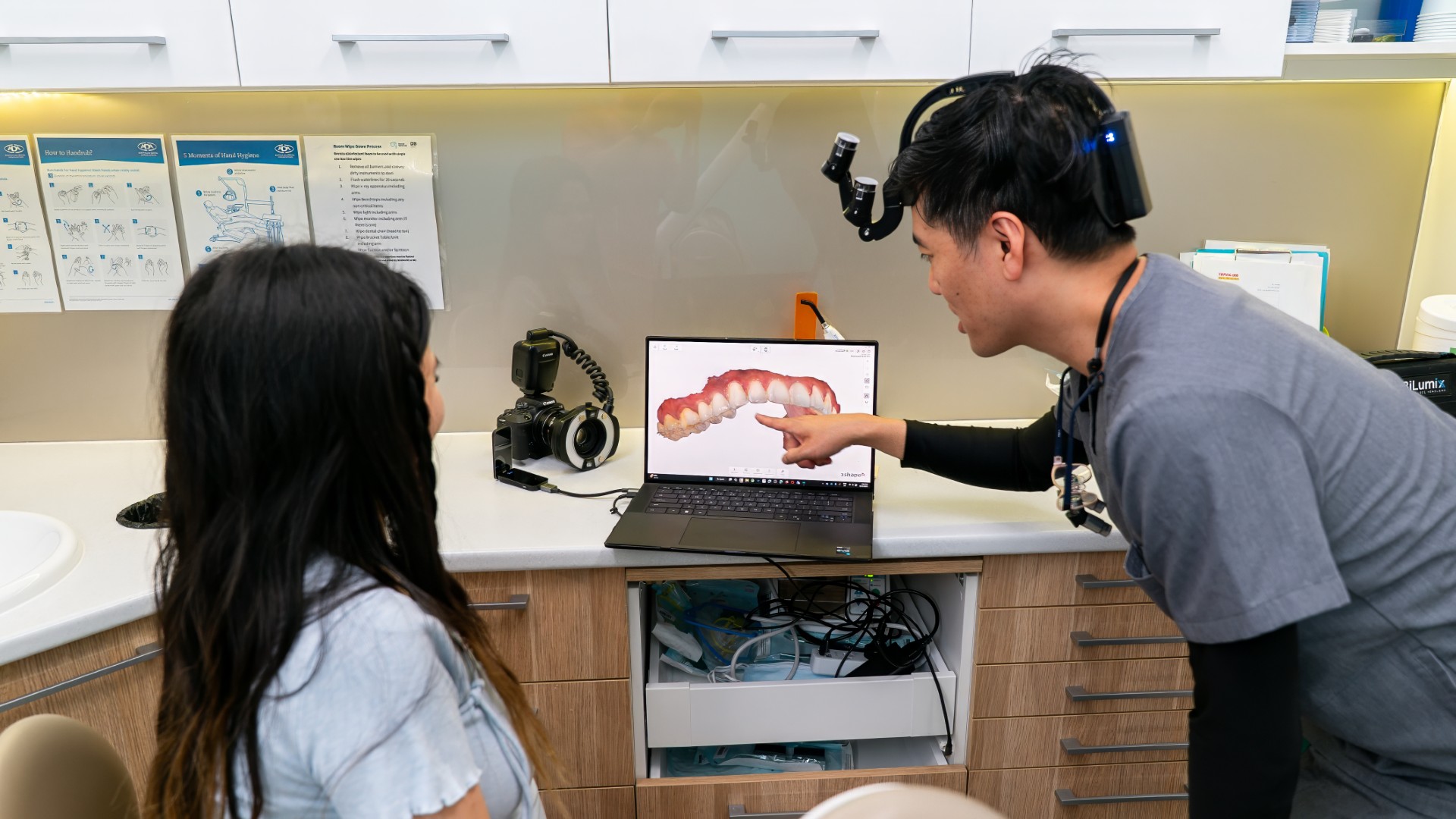
Those small telescopes on your dentist's glasses do two jobs. They make fine detail such as a filling edge or a tiny canal opening easier to see, and they let the dentist sit upright instead of bending over your mouth. The research supports both, though it is much stronger on what magnification helps a dentist find than on whether it changes how long your treatment lasts.
Loupes, microscopes and how they differ
Dental loupes are a pair of small telescopes fixed to glasses or a headband, made for one wearer and set to focus at one working distance. Because the focus is fixed, the dentist needs to keep their head at about that distance to keep the image sharp, which is why someone wearing loupes tends to stay very still and move the chair rather than themselves.
There are two common optical designs. Galilean loupes use a simple lens arrangement and are suited to lower magnifications. Prismatic loupes fold the light through prisms, which allows higher magnification with a usable field of view. Lower-power loupes show several teeth at once; higher powers show less and less of the mouth, with a shallower zone of sharp focus.
A dental operating microscope is a larger instrument mounted on an arm. It can reach higher magnification than loupes and usually has a light built into its optics. In exchange, the dentist works from a fixed position, sees a very small area at a time and generally moves more slowly. Microscopes are most associated with root canal treatment and fine surgical work.
Light matters as much as enlargement. The more an image is magnified, the darker it becomes, so loupes are often paired with a small headlight. Because that light sits close to the dentist's line of sight, it reaches into the mouth with fewer shadows than an overhead lamp, which is one reason you are offered tinted glasses.
Where magnification clearly helps: finding small anatomy
The best-known example involves a narrow extra canal often present in upper molars, called MB2, which is easy to miss during root canal treatment. In a clinical study of 312 upper molars treated by endodontists (Buhrley and colleagues, Journal of Endodontics, 2002), the canal was found in 57.4 per cent of cases when a microscope was used, 55.3 per cent with loupes and 18.2 per cent with no magnification. In upper first molars alone, the figures were 71.1, 62.5 and 17.2 per cent. Using any magnification made a large difference; the gap between loupes and the microscope was not statistically significant.
A later clinical study of 150 upper first molars (Das and colleagues, Contemporary Clinical Dentistry, 2015) found MB2 in 36 per cent of teeth by direct vision, 54 per cent with an operating microscope and 72 per cent when the microscope was combined with careful removal of a little dentine. Magnification helps, but technique and knowledge of where to look matter as well. Missed canals are one of the reasons root canal treatment is such careful work.
Where the evidence is thinner: treatment results
Seeing more does not automatically mean a better outcome, and the research here is limited. A 2024 systematic review of loupes during tooth preparation (Costa and colleagues, Journal of Clinical and Experimental Dentistry) included 11 laboratory studies, all with dental students. Loupes significantly improved working posture but did not change the quality of the preparations.
For root canal work, a 2010 systematic review (Del Fabbro and Taschieri, Journal of Dentistry) found only three prospective studies, all on endodontic surgery, and no significant difference in outcomes between loupes, a surgical microscope and an endoscope. The authors found no comparative studies at all for standard root canal treatment and called for better trials.
Much of this research measures stand-in outcomes, such as canals found or preparations scored by examiners, and some of it is done by students on model teeth. That does not mean magnification is unhelpful. It means claims that loupes or microscopes raise success rates go beyond what has been shown.
The case for loupes is partly about posture
Dental work involves long periods in fixed, awkward positions. A 2022 meta-analysis in F1000Research (Chenna and colleagues), pooling 89 estimates, put the prevalence of musculoskeletal disorders among dental healthcare workers at 78.4 per cent, with higher odds in women. In Australia, a survey of 624 dental hygienists (Hayes and colleagues, BMC Research Notes, 2013) found problems reported in the neck by 85 per cent, the shoulder by 70 per cent and the lower back by 68 per cent.
Loupes bring the detail to the eye rather than the eye to the tooth, so the clinician can see clearly while sitting more upright, and the 2024 review above supports the posture benefit. These are mostly self-reported surveys, though, and no study has shown that adopting loupes lowers a clinician's future injury rate.
What no amount of magnification can do
Magnification shows surfaces the dentist can already see. It cannot look through enamel, gum or bone, so decay between teeth or under an existing crown still needs an X-ray. Our guides to the main types of dental X-rays and 3D CBCT scans explain what imaging shows that eyes cannot.

It also does not keep a tooth dry and clean while it is being worked on. That is the job of isolation, explained in our article on why dentists use a rubber dam. And it does not replace judgement. A fine crack may be easier to spot under magnification and good light, but it still has to be tested to know whether it is the cause of pain, as described in our piece on cracked tooth syndrome.
Higher magnification can also make appointments slower rather than faster, because the field is small and the dentist has to keep re-orienting.
What you may notice in the chair
A dentist using loupes usually sits upright, keeps their head still and adjusts the chair or your head position instead of leaning in. They often lift the loupes or sit back before speaking to you, because through the optics they can see only a small part of your mouth. There may be longer pauses while an edge is checked. None of this signals a problem.
Questions patients ask about loupes
Does my dentist need loupes to do a good job?
No. Magnification is a useful tool, but a dentist who works without it is not giving poor care because of that alone. Experience, diagnosis and careful technique matter more than the equipment.
Is a dentist with a microscope better?
Not automatically. A microscope suits particular tasks, especially complex root canal work, and comes with slower working and a narrow view. The clinical study above found no significant difference between loupes and a microscope for finding the MB2 canal.
Can loupes find cavities a dentist would otherwise miss?
They can make surface detail easier to judge, such as the edge of an old filling. Decay between teeth or under a restoration still needs an X-ray.
If you are due for a check-up, the team at Lumi Dental sees new patients in Melrose Park and can take you through anything you are unsure about. See our current offers or read about general dentistry at Lumi Dental.
This article is general information only and is not personal advice. If you have a dental problem or a question about your own treatment, please speak with a dentist who can examine you.