A dental CBCT is a 3D X-ray that should only be taken when the result is expected to change the clinical decision, not as part of a routine check-up. A small field-of-view dental CBCT delivers roughly 20 to 100 microsieverts of radiation, commonly quoted as about 40 to 100 microsieverts for a single-tooth region. For comparison, average natural background radiation is roughly 5 to 8 microsieverts per day, so a small scan sits broadly in the order of a couple of weeks of everyday background exposure. That is a modest dose, but it is not zero, and it deserves a reason.
Key Takeaways
- CBCT stands for cone beam computed tomography, and it produces a 3D volume that can be sliced in any plane.
- Unlike a flat X-ray, it lets bone width be measured as well as height, and vital structures like nerves and sinuses located precisely.
- A small field-of-view CBCT is broadly about 3 to 6 times the dose of an OPG panoramic X-ray, depending on scan mode.
- Legitimate reasons include implant planning, impacted teeth, suspected root fractures, complex or failing root canals, and jaw pathology.
- The governing principle is ALARA, as low as reasonably achievable, meaning the scan must be expected to change the decision.
- A CBCT is not a routine check-up scan and should not be taken just to have a look.
What a CBCT Actually Does
A standard dental X-ray, whether a small bitewing or a wide panoramic, produces a flat image. Everything in the path of the beam is compressed into two dimensions. That works well for detecting decay between teeth and checking bone levels, but it hides depth entirely. Two structures sitting one behind the other overlap in the picture, and you cannot tell which is in front.
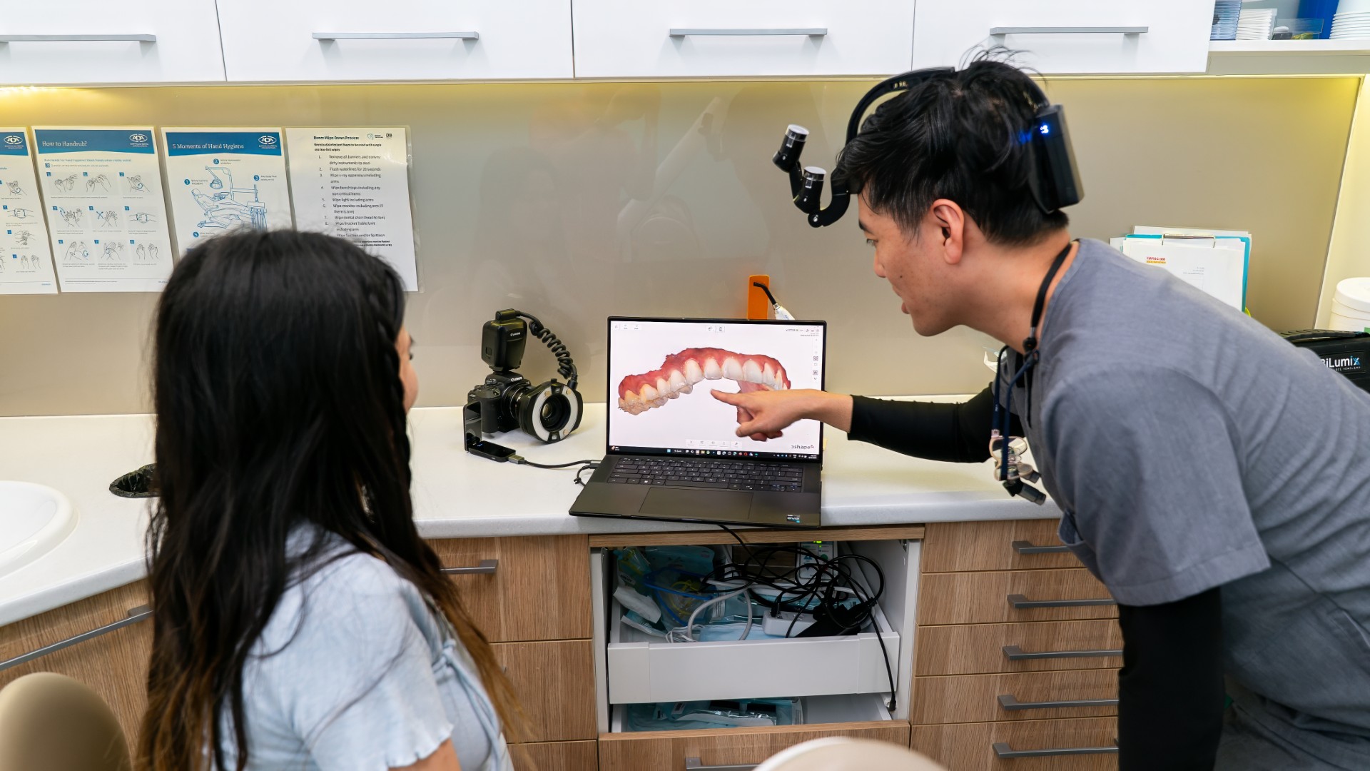
A CBCT machine rotates a cone-shaped beam around your head and captures a series of images, which software reconstructs into a 3D volume. Your dentist can then scroll through that volume slice by slice, in any direction, and take measurements. That difference is not cosmetic. It answers questions a flat film physically cannot.
The Three Questions Only 3D Can Answer
First, how wide is the bone? A flat film shows you height only. An implant needs bone around it in every direction, so width matters as much as height. Second, exactly where does the nerve run? The inferior alveolar nerve in the lower jaw and the maxillary sinus above the upper back teeth both need to be located in three dimensions before surgery near them. Third, what is the shape of this root or canal? Extra canals and unusual curvature are common and frequently invisible on a 2D film.
Radiation, Stated Plainly
Most pages about CBCT are written by clinics that own a scanner, and radiation gets a single reassuring sentence. Here are the numbers so you can judge for yourself.
| Scan type | Typical dose | Roughly equivalent to |
|---|---|---|
| Single bitewing or PA X-ray | Around 5 to 9 microsieverts (one reference gives a PA at 8.90) | Roughly one to two days of natural background radiation |
| OPG panoramic | Roughly 15 to 22 microsieverts (one reference gives 21.87) | Roughly two to four days of natural background radiation |
| Small field-of-view dental CBCT | Roughly 20 to 100 microsieverts, commonly 40 to 100 for a single-tooth region | Roughly one to two weeks of natural background radiation |
| Medical CT of the head | Substantially higher again, in the order of a thousand or more microsieverts | Many months of natural background radiation |
Average natural background radiation is roughly 5 to 8 microsieverts per day, which is the comparator used in the right-hand column. Depending on scan mode, a dental CBCT is broadly about 3 to 6 times the dose of an OPG. For context on a full 2D series, one reference set gives a PA at 8.90, an OPG at 21.87 and a lateral cephalometric at 5.03 microsieverts, totalling 35.81 microsieverts. Doses vary considerably with machine, settings and field size, so treat these as typical figures rather than fixed values.
If you want to understand the 2D films first, our guide to bitewing and OPG X-ray types covers what each one is for, and how often check-up X-rays are needed explains the intervals.
When a CBCT Is Genuinely Justified
Dental Implant Planning
This is the most common legitimate reason. Before placing an implant, the surgeon needs to know the width and height of available bone, its density, and the exact position of the inferior alveolar nerve in the lower jaw or the floor of the maxillary sinus in the upper. Getting that wrong has serious consequences, including lasting numbness. Our articles on bone grafts and sinus lifts and numbness after implant surgery explain why this planning matters.
Impacted Teeth
An impacted canine or wisdom tooth needs to be located in three dimensions before anyone operates. A CBCT shows whether an impacted canine is sitting against the root of the tooth next door and causing resorption, or whether a lower wisdom tooth root is genuinely wrapped around the nerve canal rather than just overlapping it on the flat film. Our guide to impacted canine teeth covers this situation in more detail.
Suspected Root Fracture or an Unexplained Symptom
A vertical root fracture is notoriously hard to see on a 2D film, because the fracture line often runs in the same plane as the beam. Where a tooth remains persistently symptomatic and the standard films look normal, a small CBCT may show the fracture or the characteristic bone loss pattern around it. Our articles on cracked tooth syndrome and tooth abscess signs describe the symptoms that lead here.
Complex or Failing Root Canal Treatment
Missed canals are a common reason root canal treatment does not settle. A CBCT can reveal an extra canal, an unusual root anatomy or a separated instrument that a flat film has not shown. This is a standard consideration before root canal retreatment.
Jaw Pathology
Cysts, lesions and other findings in the jaws need their full extent assessed, including how close they sit to nerves, sinuses and tooth roots. That is a three-dimensional question.

When to Ask Why
The ALARA principle, as low as reasonably achievable, is the standard that governs all dental radiography. Applied to CBCT it means one thing: the scan should be expected to change what happens next. If the treatment plan would be identical either way, the scan is not justified.
So it is entirely reasonable, and not at all rude, to ask two questions before any 3D scan: what specific question are we trying to answer with this, and would the plan change depending on the answer? A good clinician will have a clear reply, because they have already asked themselves the same thing.
A CBCT is not a routine check-up scan. It should not be taken simply to have a look, to screen a healthy mouth, or as an automatic add-on to a new patient examination. Standard bitewings and, where indicated, a panoramic film cover routine screening at a far lower dose. If you are unsure about a proposed plan involving significant treatment, our guide on when to get a second opinion may be useful. Asking questions is a normal part of care, and most clinicians welcome it.
General Market Cost
As a general guide to market ranges across Australia, a dental CBCT commonly runs roughly $150 to $400. It is sometimes bundled into an implant planning fee rather than billed separately. That is a general market range and not a quote. What you would pay depends on the scan size, whether a report is included and how it fits into your overall plan, so ask for it in writing before proceeding.
Common Questions
Is a dental CBCT safe?
The dose is low but not zero. A small field-of-view scan sits roughly in the order of one to two weeks of natural background radiation. It is considered a reasonable exposure when there is a clear clinical question that a 2D film cannot answer, and unjustified when there is not.
How much radiation is a CBCT compared to a normal X-ray?
Broadly about 3 to 6 times an OPG panoramic film, depending on the scan mode and field size, and considerably more than a single bitewing. It is still far less than a medical CT scan of the head.
Do I need a CBCT for a dental implant?
For most implant cases a CBCT is standard practice, because bone width and the position of nerves and sinuses cannot be measured reliably any other way. It is one of the clearest examples of a scan that genuinely changes the plan.
Can I have a CBCT while pregnant?
Non-urgent imaging is generally deferred during pregnancy. If imaging is clinically necessary it can usually be done safely with appropriate protection, but the decision should be made with your dentist and your doctor. Our article on dental X-ray safety during pregnancy covers this in more detail.
How long does a CBCT scan take?
The scan itself typically takes under a minute, often around 10 to 40 seconds of rotation depending on the machine and settings. You stand or sit still with your head positioned in the unit. There is no injection and nothing goes in your mouth.
Should children have CBCT scans?
Children are more sensitive to radiation, so the threshold for justifying any scan is higher. CBCT in children is generally reserved for specific situations such as an impacted tooth, a suspected pathology or complex orthodontic planning. Our guide to dental X-rays for kids explains the general approach.
This article is general information only and is not a substitute for personal dental advice. Whether any imaging is appropriate for you depends on an assessment by a registered dental practitioner.
If you have been offered a 3D scan and want to understand why, or you would like your imaging explained properly rather than glanced at, that is a fair thing to ask for. The team at Lumi Dental in Melrose Park is happy to walk you through what your images show and what they mean for your options, with a written quote after assessment. See our current deals page for what is available, or read more about care from our general dentist in Melrose Park.