
On a healthy upper central incisor, the highest point of the gum margin usually sits about 1 mm off the tooth's centre line, towards the side further from the middle of the face. Dentists call that high point the gingival zenith. It is a small detail, and studies suggest people who are not dentists tend to notice it only when the two sides of the smile differ by around a millimetre or more, but it matters when front teeth are being restored, straightened or reshaped.
Each front tooth has its own zenith position
Look closely at the gum around an upper front tooth and it forms a curve, like a shallow arch. The top of that arch is the zenith. On most incisors it is not in the middle of the tooth but slightly off centre, towards the side further from the midline of the face (dentists call this side distal).
The most quoted measurements come from Chu and colleagues (Journal of Esthetic and Restorative Dentistry, 2009), who measured 240 sites in 20 healthy adults without gum disease, crowding or fillings on these teeth:
| Tooth | Where the zenith sat (Chu 2009) | How often it was off centre (Martins 2025 review) |
|---|---|---|
| Central incisor | Distal on every tooth measured, about 1 mm on average | Distal in 96% |
| Lateral incisor | About 0.4 mm off centre on average | Distal in 84% |
| Canine | Centred on the long axis in 97.5% | Distal in 43%, centred in 44% |
A 2025 systematic review and meta-analysis in the Journal of Prosthetic Dentistry (Martins and colleagues) pooled cross-sectional studies of healthy adults and found the same pattern: the further forward the tooth, the more often and the further the zenith sits distally. The authors rated the certainty of the evidence as very low, so these figures describe a common pattern rather than a rule every smile should follow.
The lateral incisor usually sits a little lower
If you draw a line joining the zeniths of the central incisor and the canine, the gum on the lateral incisor between them usually peaks slightly below that line, closer to the biting edge. Chu and colleagues measured this at about 1 mm, and the Martins review found the lateral zenith sat below the line in 82% of people. The result is a gentle high, low, high rhythm across each side of the smile.
The Martins review also found that the left and right sides were usually symmetrical with each other. The perception studies below suggest that a difference between the two sides may matter more to how a smile looks than the exact position of any one zenith.
How much people notice
Several studies have shown people photographs of the same smile with the gum line digitally altered.
- Nomura and colleagues (Dental Press Journal of Orthodontics, 2018) changed the zeniths of the central and lateral incisors in steps of 0.5 to 1 mm. Asymmetric changes were rated less attractive than symmetric ones, differences greater than 1 mm were noticeable, and changes on the central incisors were easier to see than changes on the laterals. Lay raters only picked up the change at 1 mm of asymmetry on a central incisor, while dentists and orthodontists were more critical.
- Batra and colleagues (American Journal of Orthodontics and Dentofacial Orthopedics, 2018) asked 100 lay people to rank 67 digitally altered versions of one smile photograph. Black triangles between the teeth and changes in gum colour were ranked most negatively; changes in gum contour and zenith had the least effect. For some features, such as gum colour and the height of the gum margin, lay raters were more bothered by a change on one side than by the same change on both sides.
- Masoumi and colleagues (Journal of Dentistry, Shiraz, 2025) found that every group of raters scored the unaltered smile highest and an asymmetric zenith change lowest, but the difference was statistically significant only in the three dental groups (periodontists, restorative dentists and general dentists), not among the 15 non-professionals, and the dental groups differed in how much the changes mattered to them.
Taken together, a zenith that is a fraction of a millimetre away from the textbook position is probably not the main reason a smile looks off. A clear difference between one central incisor and the other is more likely to be seen. In the Batra study, gaps at the gum between teeth stood out more than changes to the zenith, a topic covered in our guide to black triangles between teeth.
Why the zenith matters when teeth are restored
The gum margin frames the tooth. The Martins review notes that the zenith influences where the line angles of a crown appear to start as the tooth emerges from the gum and how upright or tilted the crown looks. If a veneer or crown is shaped without regard to where the gum peaks, a straight tooth may look as if it leans.
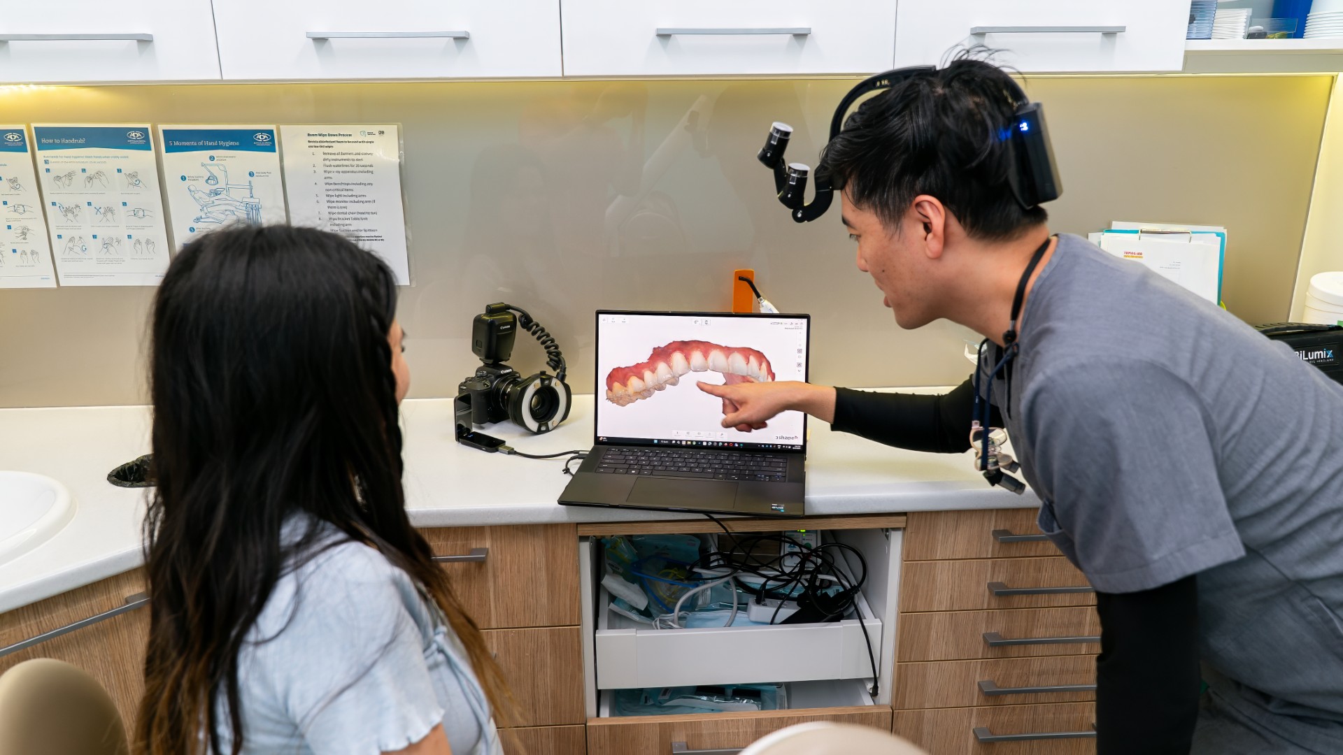
For that reason, the gum line is recorded alongside the teeth when a smile is planned. At Lumi, photographs taken in our clinical photography room and a 3D scan of the teeth are used together in digital smile planning, alongside other proportions such as the smile arc. Any digital preview is a planning tool, not a promise of the final result.

What can change where the gum peaks
The position of the gum is closely linked to the position of the tooth and the bone beneath it, so the options depend on the cause.
- Moving the teeth. When a tooth is tipped, rotated or sitting higher than its neighbour, the gum line usually moves with it. In a study of photographs from 150 patients taken before and after fixed braces, Seehra and colleagues (European Journal of Orthodontics, 2025) found overall gum appearance scores improved after treatment, and the authors concluded braces can have a positive effect on features including the zenith, although ratings varied considerably between the five assessors.
- Reshaping the gum or bone. Where the gum covers more of a tooth than usual, gum contouring or aesthetic crown lengthening may be considered. Chu and colleagues suggested their measurements could guide these procedures. Our articles on gum contouring and crown lengthening and the bone level explain what is involved.
- Restoration shape. When the gum position is acceptable, a veneer, crown or bonding can be shaped so its outline follows the existing zenith.
A healthy gum comes first. Inflammation and gum recession change the gum line too, so a dentist or OHT will usually check gum health before any cosmetic planning.
Questions about the gum line and smile design
Do both sides need to match exactly?
Few natural smiles are perfectly symmetrical. The studies above suggest small differences often go unnoticed by people who are not dentists, while a difference of about a millimetre or more between the two central incisors is more likely to be seen.
Can veneers fix an uneven gum line?
Veneers change the teeth, not the gum. They can be shaped to work with the gum line you have, but if the gum itself sits at clearly different heights, moving the teeth or reshaping the gum is usually what changes it. That is discussed at the planning stage, and our guide to gum display at rest and in a full smile covers related measurements.
Can I plan this from interstate?
Yes. You can send photographs first, then have a video or phone call before booking. Our guide to travelling to Sydney for dental treatment explains how that works.
Talking it through
If the gum line is something you notice in photos, a free cosmetic consult at Lumi Dental in Melrose Park is a chance to look at it with your teeth, gum health and goals together. You can read more about our cosmetic dentistry or see the current deals page.
Sources
- Chu SJ, Tan JH, Stappert CF, Tarnow DP. Gingival zenith positions and levels of the maxillary anterior dentition. J Esthet Restor Dent. 2009;21(2):113-20. PubMed 19368601
- Martins FV, Mattos de Santana CM, Magno MB, et al. Gingival zenith level, position, and symmetry: a systematic review and meta-analysis. J Prosthet Dent. 2025;134(5):1603-1618. PubMed 38704322
- Nomura S, Freitas KMS, Silva PPCD, et al. Evaluation of the attractiveness of different gingival zeniths in smile esthetics. Dental Press J Orthod. 2018;23(5):47-57. PubMed 30427493
- Batra P, Daing A, Azam I, et al. Impact of altered gingival characteristics on smile esthetics: laypersons' perspectives by Q sort methodology. Am J Orthod Dentofacial Orthop. 2018;154(1):82-90.e2. PubMed 29957325
- Masoumi M, Rashidi Maybodi F, Sabet M, Farahat F. Effects of shape and position of gingival margin of maxillary anterior teeth on perception of smile aesthetics by periodontists, restorative dentists, general dentists, and non-professionals. J Dent (Shiraz). 2025;26(3):274-283. PubMed 40893984
- Seehra J, DiBiase AT, Alani A, et al. The effect of fixed orthodontic appliances on gingival aesthetics of the anterior maxillary dentition. Eur J Orthod. 2025;48(1):cjaf108. doi:10.1093/ejo/cjaf108. PubMed 41479362
This article is general information only and is not a substitute for advice about your own situation from a dentist.