Your dentist points to a bright white patch in the jawbone on your X-ray, often near the roots of a lower back tooth, and says it is probably nothing to worry about. Dense bone blocks more X-rays, so it shows up whiter than the bone around it. Two common explanations are a dense bone island, which is usually regarded as a variation of normal bone, and condensing osteitis, which is bone reacting to long-standing irritation from a tooth's nerve. One usually needs no treatment, while the other points to a tooth that may.
Dense bone islands are common and usually harmless
Dense bone islands go by several names, including idiopathic osteosclerosis and enostosis. They are usually found by chance on X-rays taken for other reasons and usually cause no symptoms.
How often they are seen depends on the population and the type of X-ray. In a study of 2,504 people who had 3D cone beam scans at a Beijing hospital, 13.9% had at least one bone island, most often in the premolar and molar region of the lower jaw (Zhou and colleagues, BMC Oral Health, 2025). On panoramic X-rays, a Lebanese study of 520 adults found idiopathic osteosclerosis in 9% (Hayek and colleagues, Medical Archives, 2023), and a study of 571 orthodontic patients in China found it in 11.3%, all in the lower jaw (Wang and colleagues, Journal of Dental Sciences, 2022).
A Norwegian study followed 210 dental school patients with X-rays for 10 to 17 years, and 130 of them for a further 10 years. Sixteen had idiopathic osteosclerosis, mostly in the lower molar and premolar region. Over that time a few lesions shrank, one disappeared and a few new ones appeared, and the authors concluded that these lesions should be considered anatomical variants (Halse and Molven, International Endodontic Journal, 2002).
They can grow in teenagers and then settle
Bone islands are not always fixed in size. In the study of orthodontic patients, 68 lesions were followed for an average of 36 months. About 56% grew, 10% shrank and 34% stayed the same. Growth was most common in people aged 10 to 19, where 76.5% of lesions enlarged, while 80% of lesions in people aged 30 to 39 did not change (Wang and colleagues, 2022). The authors suggested that the lesions develop early in life and become fairly stable in adulthood. So a bone island that grows during the teenage years is not unusual, though growth is something your dentist may note.
Condensing osteitis is a reaction to a problem in the tooth
Condensing osteitis looks similar but has a cause. It is a dense area of bone around the root tip that forms in response to chronic low-grade inflammation coming from the tooth's nerve (the pulp). A 2026 meta-analysis of 10 studies including 17,285 people estimated its prevalence at about 3.9%, although the studies varied a great deal and the certainty of that figure was rated very low (Segura-Egea and colleagues, International Endodontic Journal, 2026). In the Lebanese study, condensing osteitis was seen on 5.8% of panoramic X-rays, and the authors linked it to pulp inflammation, deep fillings and decay (Hayek and colleagues, 2023).
Because it reflects a problem in the nerve, the tooth involved often has a deep filling or decay, and the nerve may be inflamed or dying. The more familiar sign of nerve infection is a dark area at the root tip, which our article on dark areas at the root tip explains. Condensing osteitis can be thought of as the bone responding in the opposite direction.
How a dentist tells them apart
No single feature settles it, so the dentist or OHT looks at the X-ray and the tooth together.
| Feature | Dense bone island | Condensing osteitis |
|---|---|---|
| Relationship to the tooth | Can sit well away from the teeth, or near the roots of healthy teeth | Around the root tip of a tooth with a nerve problem |
| The tooth nearby | Usually healthy, with a nerve that responds normally | Often has deep decay or a large filling, with an inflamed or infected nerve |
| Space around the root | The thin dark line around the root (the ligament space) can usually be seen clearly | This space was widened on 38 of 49 roots in one series before treatment |
| Symptoms | Usually none | Often none |
| What usually happens | Recorded and left alone | The tooth is treated, usually with root canal treatment or removal |
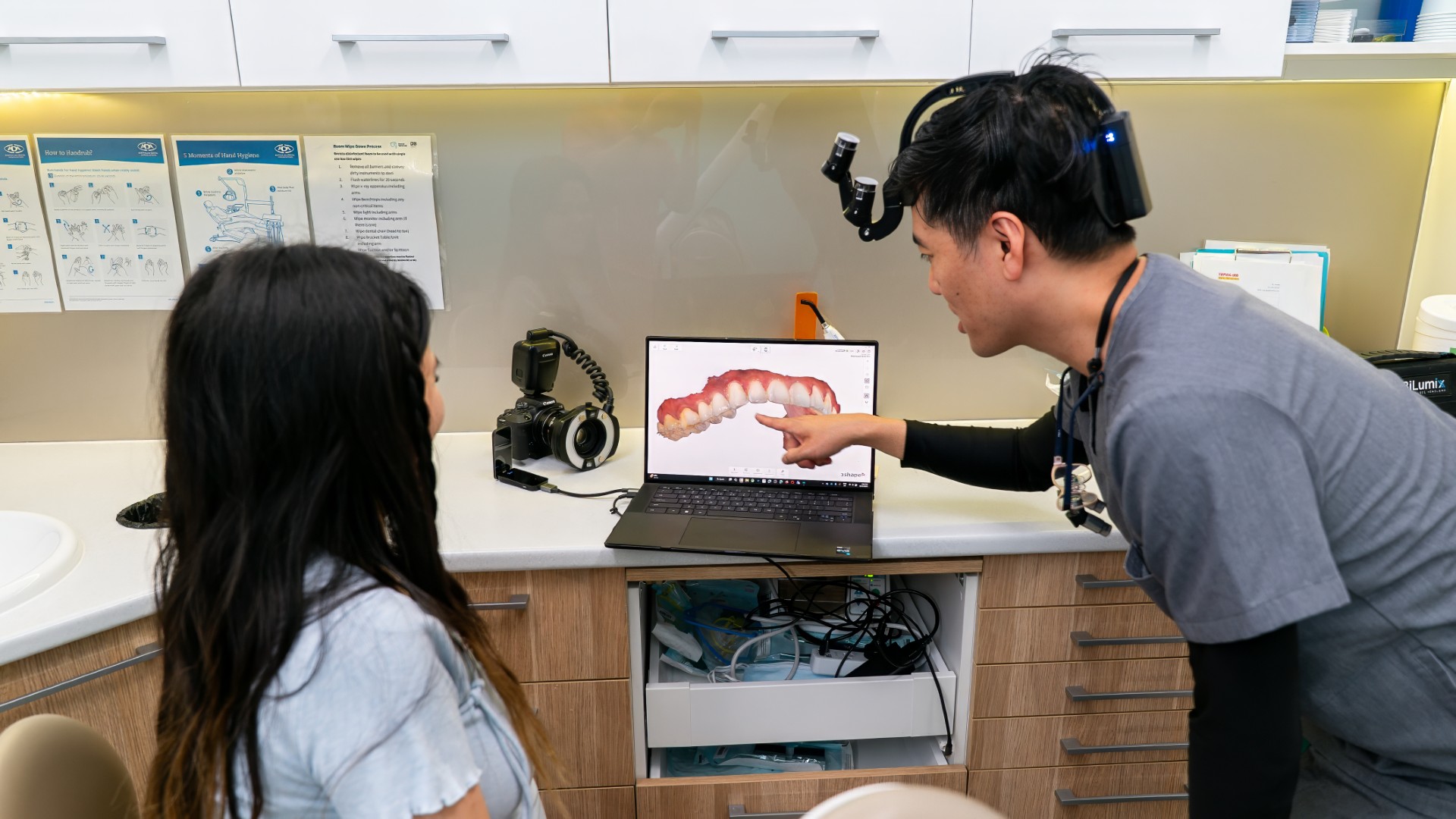
The sources for this table are the diagnostic criteria used by Wang and colleagues (2022) and Zhou and colleagues (2025), the description in Hayek and colleagues (2023), and the treatment series by Eliasson and colleagues (1984). Tests of the nerve, such as a cold test, help, but they are not perfect: in a meta-analysis, the cold test had a pooled sensitivity of 0.87 and specificity of 0.84 (Mainkar and Kim, Journal of Endodontics, 2018). A 3D X-ray can show where a dense area sits in relation to the root, which is how the Beijing study classified bone islands. Lumi's X-ray unit can take both panoramic and 3D images.

Condensing osteitis often fades after the tooth is treated
A study followed 49 roots of 36 teeth with condensing osteitis for an average of 4.3 years after root canal treatment. The dense bone had disappeared completely around 36 of the 49 roots, and none had progressed. The widened ligament space seen on 38 roots before treatment remained on only 12 at follow-up (Eliasson and colleagues, Oral Surgery, Oral Medicine, Oral Pathology, 1984). This is an old and small study, but it supports the idea that the bone change follows the tooth: treat the source of the irritation and the bone tends to return to a normal appearance. healthdirect has a plain language guide to root canal treatment, and our root canal treatment guide covers what is involved.
When a white patch needs a closer look
Most dense areas fall into one of these two groups, but a few findings prompt further questions.
- Several dense areas or bony growths. Multiple bone islands or bony growths called osteomas can be a feature of Gardner syndrome, an inherited condition linked with bowel polyps, so a dentist may consider this possibility (Wang and colleagues, 2022; Zhou and colleagues, 2025).
- A lesion that expands or moves teeth. A dense area that swells the bone, displaces teeth or keeps growing raises the possibility of an osteoma or another lesion, and a tissue sample may be needed to confirm it (Wang and colleagues, 2022).
- Patches at the tips of several healthy lower teeth. Cemento-osseous dysplasia, a condition of the bone around the roots, can be mistaken for infection. In one reported case, the teeth responded to sensitivity testing, which pointed away from root canal treatment (Delai and colleagues, Journal of Endodontics, 2015).
Our article on bony lumps in the mouth covers a different kind of bony growth. Our article on pulp stones covers white spots inside the tooth itself.
Bone islands and braces
Evidence on whether bone islands get in the way of orthodontic treatment is limited and mixed. A case report and review discussed possible difficulty closing spaces or achieving the right root angle (Sinnott and Hodges, Journal of Orthodontics, 2020). In the study of 571 orthodontic patients, however, the authors found no hindrance to tooth movement and no root resorption around the lesions, while noting the limits of panoramic X-rays (Wang and colleagues, 2022).
For dentists: test the tooth before treating the radiopacity
Before planning endodontic treatment for a tooth next to a periapical radiopacity, check the tooth as well as the image. Condensing osteitis is linked with pulpal disease, so a sensibility test, the state of the crown (deep caries or a large restoration) and the periodontal ligament space all help. In the Eliasson series, the ligament space was widened on 38 of 49 roots before treatment and on only 12 at follow-up. A tooth that responds normally, with an intact ligament space and no deep restoration, points towards idiopathic osteosclerosis or another non-endodontic cause. The cemento-osseous dysplasia case reported by Delai and colleagues illustrates the principle: the periapical lesions there were radiolucent rather than dense, but the teeth responded to sensitivity testing and root canal treatment was avoided.
The limits: cold testing has a pooled sensitivity of 0.87 and specificity of 0.84 (Mainkar and Kim, 2018), so one test should not decide the diagnosis, and diagnostic criteria for condensing osteitis vary between studies (Segura-Egea and colleagues, 2026). Where the relationship to the root is unclear, a limited-field 3D scan or a review against older X-rays may help.
Common questions
Do I need treatment for a bone island?
Usually not. Dense bone islands usually cause no symptoms and do not need treatment (Sinnott and Hodges, 2020), and the Beijing study concluded that they usually only need careful diagnosis (Zhou and colleagues, 2025). Your dentist may note its size so it can be compared on future X-rays.
Can condensing osteitis go away?
It often does once the tooth is treated. In the Eliasson follow-up study, the dense bone fully regressed around most roots after root canal treatment.
Is a white patch on my X-ray the same as a dark patch?
No. A dark area at the root tip usually means bone has been lost around an infected root, while a white patch means the bone is denser than usual. Both can be linked to a tooth's nerve, but a white patch can also be a harmless bone island.
Sources
- Zhou XH and colleagues. Cone-beam computed tomography assessment of the prevalence and distribution of bone islands of the jaws in a group of Chinese patients. BMC Oral Health, 2025. PubMed 41013412
- Hayek E and colleagues. Idiopathic osteosclerosis and condensing osteitis in a sample of the Lebanese population: a digital panoramic based study. Medical Archives, 2023. PubMed 38299083
- Wang S and colleagues. Longitudinal investigation of idiopathic osteosclerosis lesions of the jaws in a group of Chinese orthodontically-treated patients using digital panoramic radiography. Journal of Dental Sciences, 2022. PubMed 35028028
- Halse A and Molven O. Idiopathic osteosclerosis of the jaws followed through a period of 20 to 27 years. International Endodontic Journal, 2002. PubMed 12449025
- Segura-Egea JJ and colleagues. Radiographic prevalence of condensing osteitis: a systematic review and meta-analysis. International Endodontic Journal, 2026. PubMed 42508034
- Eliasson S, Halvarsson C and Ljungheimer C. Periapical condensing osteitis and endodontic treatment. Oral Surgery, Oral Medicine, Oral Pathology, 1984. PubMed 6583626
- Mainkar A and Kim SG. Diagnostic accuracy of 5 dental pulp tests: a systematic review and meta-analysis. Journal of Endodontics, 2018. PubMed 29571914
- Delai D and colleagues. Florid cemento-osseous dysplasia: a case of misdiagnosis. Journal of Endodontics, 2015. PubMed 26410152
- Sinnott PM and Hodges S. An incidental dense bone island: a review of potential medical and orthodontic implications of dense bone islands and case report. Journal of Orthodontics, 2020. PubMed 32419572
- healthdirect. Root canal treatment (last reviewed February 2025). healthdirect.gov.au
If something on your X-ray has been flagged and you would like it explained, you can book an appointment at our Melrose Park practice. Our general dentistry team can go through the images with you.
This article is general information only and is not a substitute for an in-person assessment by a dental professional.