Many people only learn they have a problem at the root of a tooth when a dentist points to a dark patch on an X-ray. The tooth may feel normal. Often that dark patch is a sign of apical periodontitis: inflammation at the root tip caused by bacteria inside the root canal, which leads to loss of the bone around the root. It is common, it often causes no symptoms, and in most treated teeth the lesion gets smaller once the infection inside the tooth is treated.
This article explains why the patch appears, why it is not always an infection, what treatment usually involves, and how long the bone can take to fill back in.
The dark area is lost bone, not a hole in the tooth
A 2004 review by Nair describes apical periodontitis as the body's defence response to bacteria coming from the root canal system. Where the infected root canal meets the tissue at the root tip, this response causes local inflammation and breakdown of hard tissue, including bone. Bone that has been broken down shows up as a darker area on an X-ray.
Healthdirect explains that when a tooth is damaged, bacteria can get inside it, which can lead to an infection or abscess in the root. It also notes that an untreated root canal infection can lead to chronic inflammation and damage to the tissue around the tooth, and that antibiotics alone do not work to cure root canal infections.
It is more common than many people realise
A 2021 systematic review by Tibúrcio-Machado and colleagues in the International Endodontic Journal pooled 114 studies covering 34,668 people. About half of the people studied (52%) had at least one tooth with apical periodontitis, although only about 5% of all teeth were affected. The figure was 40% in samples drawn from the general population and higher in samples from dental services and hospitals. It was much higher in teeth that had already had root canal treatment (39%) than in untreated teeth (3%). The authors noted that the condition often causes no symptoms, and that the studies varied widely and many had a high risk of bias, so these are broad estimates.
Why a tooth with a dark patch may not hurt
Healthdirect notes that a root canal infection may not always cause symptoms right away. When symptoms do appear, they can include toothache or sensitivity, swelling near the tooth or jaw, a tooth that turns darker, or a pimple-like bump near the tooth, which healthdirect describes as pus building up. Our article on toothache that stopped on its own looks at what it can mean when pain goes away, and our post on gum boils covers bumps on the gum.
No pain does not mean nothing is happening. Healthdirect advises that without treatment, the infection may get worse and the tooth may need to be removed.
Not every dark spot near a root is an infection
Other conditions can look similar on an X-ray. One that is regularly confused with infection is periapical cemento-osseous dysplasia, a benign bone condition. A 2019 report by Brody and colleagues describes cemento-osseous dysplasia as linked to teeth with a living pulp, usually without symptoms, and the periapical form as affecting the root tips of the lower front teeth. It generally does not need treatment, although follow-up is recommended. A 2015 case report and review by Senia and Sarao followed a lesion of this type under a lower incisor for 12 years, and concluded that careful pulp testing helps avoid misdiagnosis and unnecessary treatment.
Less often, a lesion in this position has nothing to do with the tooth. A 2014 case series and literature review by Sirotheau Corrêa Pontes and colleagues in the Journal of Endodontics analysed 56 lesions that had been misdiagnosed as tooth infections. Of these, 37 were benign, 16 were malignant and 3 were Stafne bone cavities. The most commonly reported benign lesions were ameloblastomas and nasopalatine duct cysts. These were selected cases, so they show what can happen rather than how often it happens. The authors recommended combining the clinical and X-ray findings with the medical history, pulp tests and aspiration, and taking a biopsy when these point away from a tooth infection.
Both papers stress testing whether the tooth still responds before treatment. A dark patch at the root tip of a tooth that still responds needs a closer look before any treatment.

A 3D scan can show what a flat X-ray misses
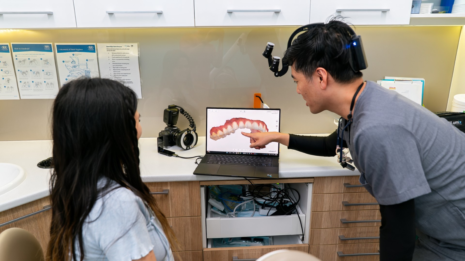
In a 2015 clinical study by Davies and colleagues of 100 root-filled teeth referred for retreatment, lesions were seen at 41% of 209 roots on a single standard X-ray and at 68% on a cone beam CT (3D) scan. A 2026 systematic review by Segura-Egea and colleagues found that studies using 3D scans reported more lesions in root-filled teeth than most single two-dimensional methods. Combining a panoramic and a standard X-ray gave estimates similar to 3D scans, and the certainty of the evidence was low to very low.
The European Society of Endodontology has published a position statement giving guidance on when a 3D scan may be used in root canal care, noting studies showing it can help with treatment planning and when reviewing treated teeth. Lumi Dental has an Orthophos 3D X-ray unit, which takes both panoramic images and 3D scans, and our article on what a dental X-ray shows covers the different types.
Treatment removes the source so the bone can repair
Nair's review describes treatment of apical periodontitis as removing or substantially reducing the bacteria in the root canal and preventing re-infection with a root filling. Healthdirect explains that root canal treatment aims to clean, disinfect and seal the root canal system. Our article on vital pulp therapy explains when treatment that tries to keep the pulp alive may be considered instead.
Options a dentist may discuss include:
- root canal treatment, when the tooth has enough structure left to be restored
- retreatment, when a tooth that already has a root filling shows a lesion that has not healed or has appeared later
- removing the tooth, which healthdirect notes is a choice instead of root canal treatment
Our guides to root canal treatment and root canal retreatment cover each option.
Most lesions shrink after treatment, but the bone takes time
In a 1996 study by Ørstavik in the International Endodontic Journal, root-treated teeth were followed with yearly X-rays for up to 4 years. Of 126 roots with a lesion before treatment, 111 (88%) showed signs of healing. Healing most often began to show at the 1-year review, and at least 89% of healing roots showed signs of it by then, but complete healing sometimes took 4 years.
A large prospective study by Ng and colleagues in 2011, with treatment carried out by postgraduate endodontic students at a university clinic, found complete healing at the root tip in 83% of roots after a first root canal treatment and 80% after retreatment, with follow-up of 2 to 4 years. Healing was more likely when a lesion was smaller, when there was no sinus tract before treatment and when the tooth had a satisfactory final restoration. A companion paper from the same study found that about 95% of treated teeth were still in place after 4 years.
A 2024 study by Zhang and colleagues looked back at 3D scans measuring lesion volume in single-rooted teeth over 4 years. Of the 125 teeth reviewed that were still present, the lesion became smaller in 116 (90.6%). Of 43 teeth whose lesion was smaller at the 1-year scan, 42 still had a smaller lesion at 4 years.
These figures come mostly from university settings and use different measuring methods, so they describe what can happen rather than a promise for any one tooth. In these studies, follow-up X-rays were compared with the X-ray taken before treatment to see whether the dark area was getting smaller.
A good seal on top is part of the treatment
A 2008 systematic review by Ng and colleagues found four factors linked with better results after a first root canal treatment: no lesion before treatment, a root filling without voids, a root filling ending within 2 mm of the root tip on the X-ray, and a satisfactory restoration on top. The authors concluded that treatment should aim to prevent re-infection with a good quality restoration. In the 2011 tooth survival study, having a permanent cast restoration rather than a temporary one was one of the factors linked with tooth survival. Our article on crowns after root canal explains when one is advised.
When to get help quickly
Pain or swelling around a tooth needs prompt attention. Healthdirect advises emergency medical care if toothache or gum swelling causes problems breathing or speaking, or swelling around the eye or neck. It advises seeing a dentist if toothache and gum swelling come with a fever over 38°C, trouble swallowing, pain when you bite, a bad taste or swelling in the cheek or jaw, and seeing a dentist as soon as possible if toothache and gum swelling last more than 2 days or do not improve with pain relief. Our emergency dental care page explains how to arrange an emergency appointment.
For dentists: pulp test before you treat, and give healing time
Before attributing a periapical radiolucency to the tooth in front of you, consider pulp testing it and its neighbours. Periapical cemento-osseous dysplasia is associated with vital teeth and affects the anterior mandible (Brody and colleagues, 2019; Senia and Sarao, 2015), and a series of 56 lesions misdiagnosed as apical periodontitis included 16 malignant lesions (Sirotheau Corrêa Pontes and colleagues, 2014). A radiolucency on a tooth that still responds to sensibility testing is a reason to reconsider the diagnosis before access. Keep in mind that sensibility tests are not definitive: a 2018 meta-analysis by Mainkar and Kim pooled a sensitivity of 0.87 and specificity of 0.84 for cold testing, so a single result should be read with the clinical and radiographic findings.
In Davies and colleagues' study, a second periapical at a different horizontal angle identified more canals than a single parallel film, but it did not increase lesion detection (38% vs 41% of roots), while CBCT found lesions at 68% of roots. Lesion presence was judged by a consensus panel on each modality in teeth referred for retreatment, so these figures show differences in detection rather than accuracy against histology. For review, Ørstavik found at least 89% of healing roots showed change by 1 year, while complete resolution sometimes took 4 years. Zhang and colleagues found that 42 of 43 lesions smaller on CBCT at 1 year remained smaller at 4 years, although that study was retrospective and limited to single-rooted teeth. A lesion that is smaller but not gone at the first review may simply need more time.
Questions we are often asked
Can the dark spot go away without treatment?
When the cause is an infection inside the root canal, healthdirect advises that without treatment the infection may get worse, and that antibiotics alone do not work to cure root canal infections. A dark area caused by something else, such as cemento-osseous dysplasia, may only need monitoring.
My tooth had a root canal years ago and now shows a dark spot. Did it fail?
Not necessarily. It may be a lesion that is still healing, since complete healing sometimes took 4 years in Ørstavik's study. It may also be a lesion that never fully resolved, or a new one: in the same study, 6% of roots with no lesion before treatment developed one. Comparing the new X-ray with older ones, if they are available, can help. Our article on a root canal that has failed covers the next steps when it has.
Is the dark area dangerous to the rest of my body?
Studies have found associations between apical periodontitis and conditions such as diabetes and cardiovascular disease. A 2019 review by Segura-Egea and colleagues explains that an association alone does not prove that one causes the other. The more direct concern is local: healthdirect notes that an untreated root canal infection can lead to chronic inflammation and damage to the tissue around the tooth.
Sources
- Tibúrcio-Machado CS and colleagues. The global prevalence of apical periodontitis: a systematic review and meta-analysis. International Endodontic Journal, 2021. PubMed 33378579
- Nair PN. Pathogenesis of apical periodontitis and the causes of endodontic failures. Critical Reviews in Oral Biology and Medicine, 2004. PubMed 15574679
- Ørstavik D. Time-course and risk analyses of the development and healing of chronic apical periodontitis in man. International Endodontic Journal, 1996. PubMed 9206419
- Ng YL and colleagues. A prospective study of the factors affecting outcomes of nonsurgical root canal treatment: part 1, periapical health. International Endodontic Journal, 2011. PubMed 21366626
- Ng YL and colleagues. A prospective study of the factors affecting outcomes of non-surgical root canal treatment: part 2, tooth survival. International Endodontic Journal, 2011. PubMed 21366627
- Ng YL and colleagues. Outcome of primary root canal treatment: systematic review of the literature, part 2. Influence of clinical factors. International Endodontic Journal, 2008. PubMed 17931388
- Zhang MM and colleagues. The trend of radiographic healing after root canal treatment in teeth with apical periodontitis based on cone-beam computed tomography: a 4-year longitudinal study. Journal of Dentistry, 2024. PubMed 38740248
- Davies A and colleagues. The detection of periapical pathoses in root filled teeth using single and parallax periapical radiographs versus cone beam computed tomography: a clinical study. International Endodontic Journal, 2015. PubMed 25074727
- Segura-Egea JJ and colleagues. Influence of imaging modality on the detection of periapical lesions in root-filled teeth: a systematic review and meta-analysis. International Endodontic Journal, 2026. PubMed 42529839
- Patel S and colleagues. European Society of Endodontology position statement: use of cone beam computed tomography in endodontics. International Endodontic Journal, 2019. PubMed 31301231
- Senia ES, Sarao MS. Periapical cemento-osseous dysplasia: a case report with twelve-year follow-up and review of literature. International Endodontic Journal, 2015. PubMed 25425097
- Brody A and colleagues. Difficulties in the diagnosis of periapical translucencies and in the classification of cemento-osseous dysplasia. BMC Oral Health, 2019. PubMed 31291935
- Sirotheau Corrêa Pontes F and colleagues. Nonendodontic lesions misdiagnosed as apical periodontitis lesions: series of case reports and review of literature. Journal of Endodontics, 2014. PubMed 24331985
- Mainkar A, Kim SG. Diagnostic accuracy of 5 dental pulp tests: a systematic review and meta-analysis. Journal of Endodontics, 2018. PubMed 29571914
- Segura-Egea JJ and colleagues. Endodontics and diabetes: association versus causation. International Endodontic Journal, 2019. PubMed 30667519
- Healthdirect Australia. Root canal treatment. healthdirect.gov.au
- Healthdirect Australia. Toothache and gum swelling. healthdirect.gov.au
This article is general information only and is not a substitute for an in-person assessment by a dental professional.