Many patients assume a mould of their teeth is only taken when something is being made, such as a crown or a mouthguard. In fact the shape of your teeth and bite is a clinical record in its own right. It helps your dentist plan treatment, track slow changes like wear or tooth movement, and talk precisely with a dental laboratory.
That shape can be captured in three related ways: a traditional impression, a study model and a digital scan. They overlap, but each has a slightly different job, and they are stored differently once treatment is finished.
Impressions come first, models come from them
An impression is the capture step. A tray loaded with a soft, putty-like material is pressed over the teeth and held still for a short time until it sets, leaving a negative copy of every surface. On its own, an impression is rarely kept. It is either sent to a laboratory to make something, such as a crown, denture, splint or mouthguard, or used to pour a model.
Impressions are an old and dependable technique. They are still the better choice in some situations, for example certain denture stages or a mouth that opens too little for a scanner to reach the back teeth comfortably.
A study model is for looking at your bite from every angle
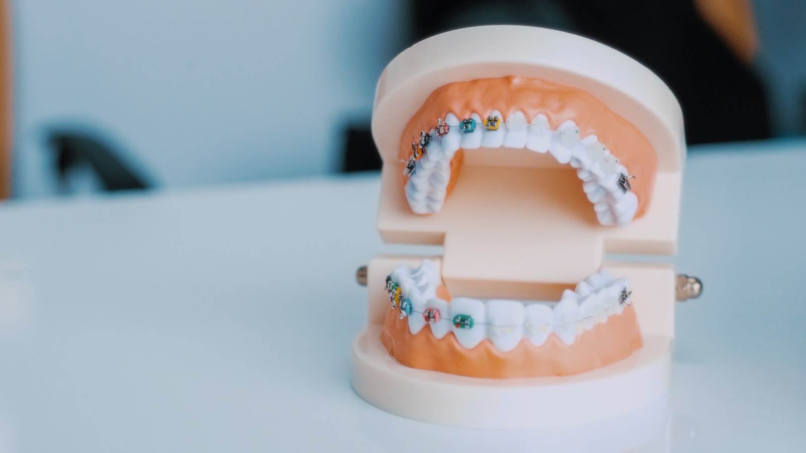
A study model is a solid replica of your teeth. Traditionally it was poured in dental stone (a type of plaster) from an impression. Today it may just as often be 3D printed from a scan. Holding a model, a dentist can look at the bite from behind and underneath, which is hard to do inside a mouth, and measure spaces and tooth positions directly.
Models are particularly helpful in a few situations:
- Orthodontic planning, where models from the start and later stages show how the teeth have moved.
- Grinding and wear, where models taken a year or two apart show whether flat spots are spreading.
- Larger rebuilding work, where upper and lower models are mounted on an articulator, a hinged device that copies how the jaws open and close, so the new bite can be planned before anything is changed in the mouth.
An intraoral scan captures the same shape as a file
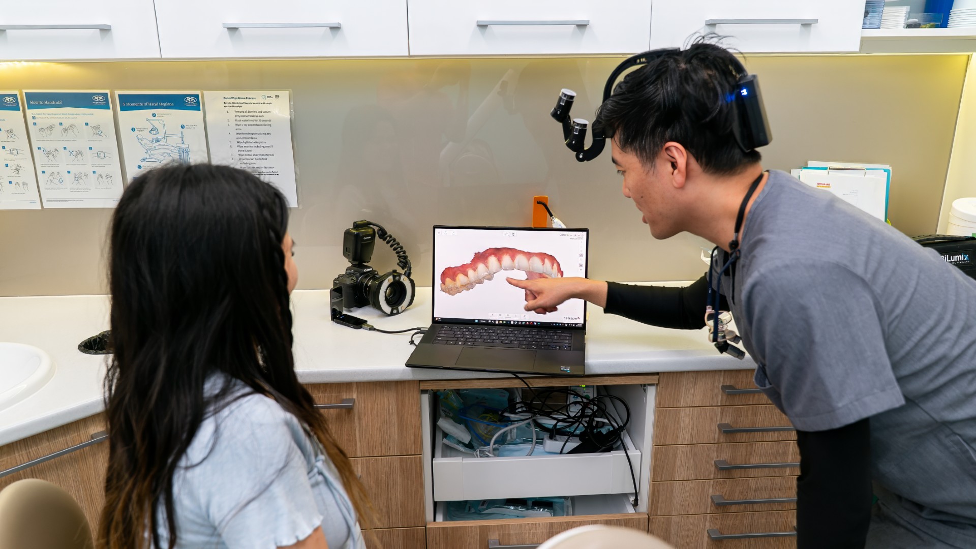
An intraoral scanner is a hand-held wand with a camera in the tip. As it passes over the teeth, software stitches thousands of images into a 3D model that appears on the screen during the scan. There is no tray to hold in the mouth, which many people find more comfortable, especially anyone who gags easily.

Because the result is a digital file, it can be sent to a laboratory electronically, enlarged on screen to check fine detail, and kept without taking up shelf space. A later scan can be laid over an earlier one to show tooth movement or wear. Scans are now routinely used for clear aligners, retainers, night guards, crowns and many other appliances.
Both methods can produce accurate results when done carefully. Which one is used depends on what is being made, how many teeth are involved and the individual mouth.
How the three compare
| Record | What it is | Typical use | What is usually kept |
|---|---|---|---|
| Impression | Set material in a tray, a negative copy of the teeth | Making a model or sending to a laboratory | Rarely kept once the model or appliance is made |
| Study model | Stone or 3D-printed replica of the teeth | Studying the bite, comparing change over time | The physical model, or a scanned copy of it |
| Intraoral scan | Digital 3D file made with a camera wand | Planning, laboratory work, comparison over time | The digital file |
Practices must keep these records for years
In New South Wales, the Health Records and Information Privacy Act requires private health service providers to keep a patient's health information for seven years from their last visit if the information was collected when they were an adult. If it was collected when they were under 18, it must be kept until they turn 25. Models and scans that form part of your clinical file fall within that record.
Digital files are easy to keep for longer than the minimum. Physical models are bulkier, so once treatment is complete and stable a practice may keep a scanned copy and return or dispose of the model itself in line with its records policy.
Getting a copy of your scan or model
You can ask for access to your health information, and that includes scans and models. A digital file can be copied without losing the original. A physical model is a single object, so a practice might offer a photograph, a scan of the model or a printed duplicate instead. If you are moving practices, our guide to changing dentists and transferring records explains how the whole file is usually transferred, and our article on who owns your dental X-rays covers the same questions for radiographs.
What patients ask about scans and models
Why take a scan when nothing seems wrong?
A baseline scan makes later comparison possible. Slow changes such as wear from grinding or gradual crowding are much easier to see by comparing two records than by memory.
Is a scan safe?
Yes. An intraoral scanner uses light to capture images, not X-rays. It records shape only, so it does not replace X-rays when your dentist needs to see inside teeth or bone. Our guide to dental X-ray types explains what each kind of image shows.
Where do models and scans fit in my treatment plan?
They are usually part of the planning records along with photos, X-rays and your examination notes. Our guide to understanding your dental treatment plan describes the other documents that usually sit in your file.
Asking about your records at Lumi
At Lumi Dental we take digital impressions with a 3D intraoral scanner and have an in-house lab. If you would like to know what records we hold for you, or want a copy for a second opinion or a new dentist, the team can explain what is available and how to request it. New patient offers are listed on our current deals page.
This article is general information about dental records in Australia and does not replace advice from the practice that currently holds your file.