When the gum around an implant crown seems to have pulled back, the mirror can be misleading. A dark shadow at the gum, a strip of metal that was not visible before, and bleeding with lost bone can all look like "receding gum", yet they range from a purely cosmetic issue to an infection that needs prompt care. A dentist separates them with a probe, a close look at the tissue and an X-ray compared with the one taken when the crown was fitted.
Written by Dr James Tran, BDent (University of Sydney), MICOI, GradDip (Oral Implants), general dentist at Lumi Dental in Melrose Park.
Inflammation around implants is common
A 2025 systematic review for the Academy of Osseointegration and the American Academy of Periodontology (Galarraga-Vinueza and colleagues, Journal of Periodontology) pooled 102 studies. It estimated that about 46% of people with implants had peri-implant mucositis, an inflammation of the gum only, and about 21% had peri-implantitis, where the bone supporting the implant is also being lost. Gum disease in the past, smoking, diabetes and alcohol were linked with a higher risk of peri-implantitis.
Recession without infection is also common. A companion review in the same journal (Tavelli and Barootchi, 2025) found gum margins moving down at many healthy implant sites, more often where the gum was thin, where the band of firm keratinised gum was narrow or missing, and after an implant was placed straight into a fresh extraction socket.
So a change at the gum line is not rare, and it is not automatically a sign the implant is failing. What matters is which of the three situations below it is.
A grey shadow with healthy gum is usually thin tissue
Gum varies in thickness from person to person. Where it is thin, the metal connector under the crown (the abutment) can show through as a grey or dark tinge. Nothing has moved and there is no infection. It is most noticeable on upper front teeth with a titanium abutment.
Abutment colour makes a difference. A 2018 meta-analysis in the Journal of Dentistry (Cai and colleagues) found less discolouration of the gum around tooth-coloured zirconia abutments than around titanium or gold-coloured ones. For a grey shadow that bothers you, the options are to leave it, to replace the abutment and crown with a zirconia one, or to thicken the gum with a soft tissue graft, which our gum graft guide describes.
A visible metal collar means the gum margin has moved
Here the gum edge sits lower on the implant than it did, so the polished collar, or occasionally a thread, is exposed and the crown may look longer. Thin gum, a narrow band of keratinised tissue, an implant positioned towards the cheek side of the bone, a crown shape that gives the gum little support, hard scrubbing and smoking can all contribute.
The implant itself can still be stable. The useful question is whether bone has been lost along with the gum, which the X-ray answers. Gum that has moved down an implant does not grow back by itself, as our article on whether receding gums can grow back explains. Treatment starts with removing the cause: a soft brush and gentler technique, help to stop smoking, and reshaping a crown that leaves the gum unsupported. When the bone underneath is sound, a connective tissue graft may cover the collar. Where grafting is unlikely to work well, a new crown designed to hide the collar is an alternative.
Bleeding, swelling or pus points to infection
The 2017 World Workshop classification (Berglundh and colleagues, Journal of Periodontology, 2018) describes the main sign of peri-implant mucositis as bleeding on gentle probing, sometimes with redness, swelling or pus. Peri-implantitis adds progressive bone loss on X-ray, often with deeper pockets and sometimes recession of the gum margin. Plaque is the driver in both.
Mucositis can usually be reversed with professional cleaning around the implant and better daily cleaning at home. Peri-implantitis is harder to treat, so catching inflammation early is worth a lot. Treatment typically begins with careful cleaning of the crown and any exposed implant surface using instruments that do not scratch titanium. Many cases then need a surgical procedure to open the gum, decontaminate the implant surface and, where the bone shape allows, graft bone. Antibiotics are sometimes added but do not replace the cleaning. Our page on peri-implantitis treatment goes into more detail, and if you had gum disease before your implant, implants and periodontitis explains why that history raises the risk.
How the three are told apart at the chair
| Situation | What you may notice | What the examination shows | Usual options |
|---|---|---|---|
| Thin gum showing the abutment | Grey or dark tinge, no soreness | No bleeding, bone level unchanged, thin tissue | Leave it, zirconia abutment and new crown, or soft tissue graft |
| Recession with an exposed collar | Metal ring visible, crown looks longer | Gum margin lower than at fitting, bone usually stable | Remove the cause, soft tissue graft, or a redesigned crown |
| Peri-implant mucositis | Bleeding when brushing, tenderness | Bleeding on gentle probing, no bone loss | Professional cleaning, home care changes, review |
| Peri-implantitis | Swelling, pus, bad taste, sometimes recession | Bleeding or pus, deeper pockets, bone loss against baseline | Cleaning, then often surgery to clean the surface and graft bone |
Probing around an implant is done gently, because there is no ligament to resist the probe and heavy pressure can cause bleeding even in healthy tissue. Depths are compared with earlier visits rather than judged against a single number. The World Workshop recommends recording X-rays and probing depths once the implant crown is completed, so that later changes can be measured against that starting point. If your implant was placed at another clinic, asking that clinic for your earlier X-rays helps.
The amount of firm, keratinised gum is also noted. The evidence on whether it protects implants long term is mixed, but the World Workshop report notes it may make the area more comfortable to brush and easier to keep free of plaque.
Looking after the gum around an implant
Daily plaque removal is the first step in managing gum disease in the Australian Dental Association's patient guidance, and the same applies to implants. Brush twice a day with a soft brush angled into the gum line. Clean between teeth with an interdental brush sized to the gap, or with superfloss where the crown sits tightly against its neighbours; our guide to floss threaders and superfloss shows how. Not smoking helps both the gum and the bone.
Regular maintenance visits matter as much as home care. At these visits the gum is probed, plaque and deposits are removed, and an X-ray is repeated when due. Your dentist will set how often you need them based on your gum history and risk; our implant aftercare guide covers the routine. Bleeding, swelling or a bad taste between visits is a reason to contact us sooner rather than waiting.
Implant gum checks at Lumi Dental
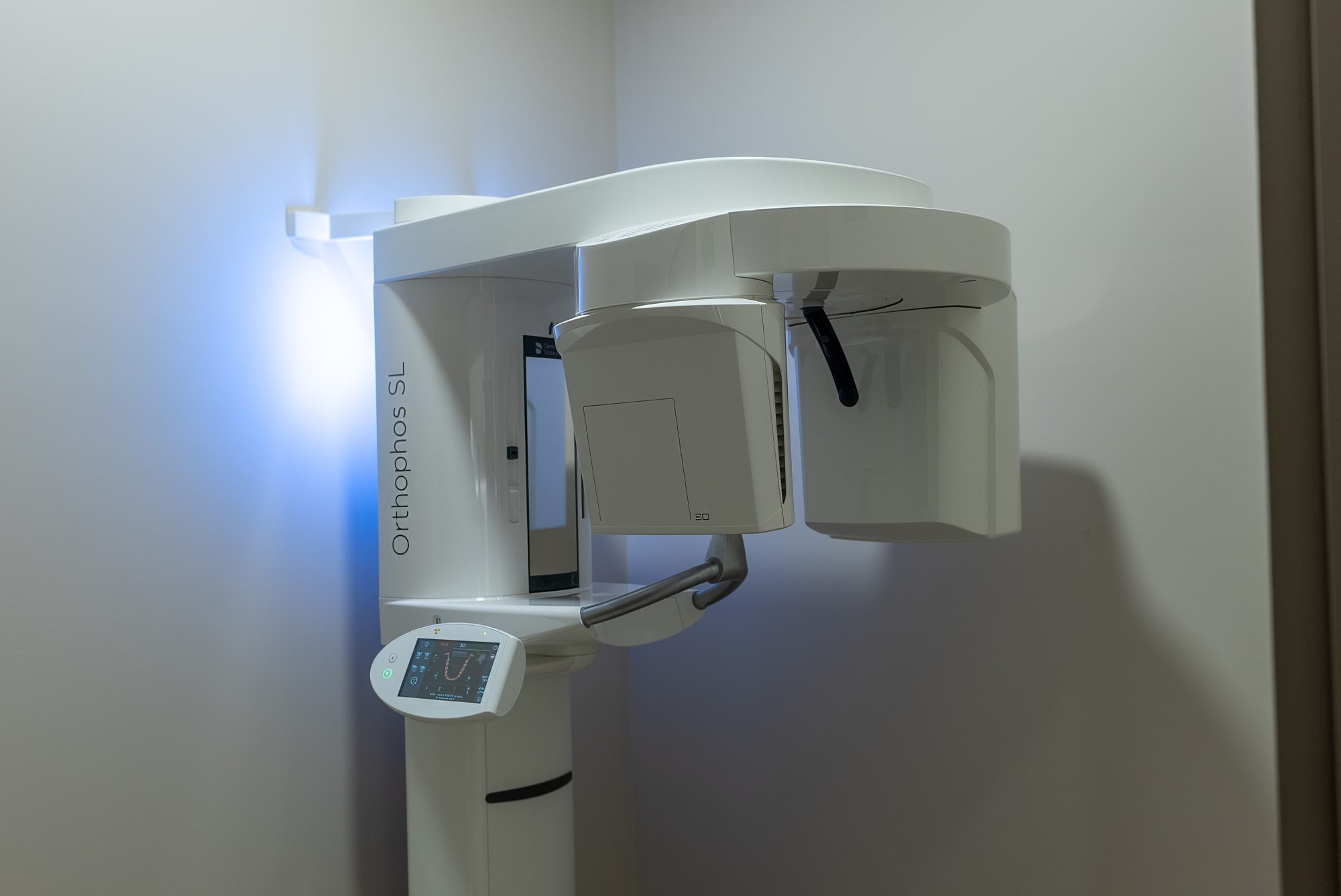
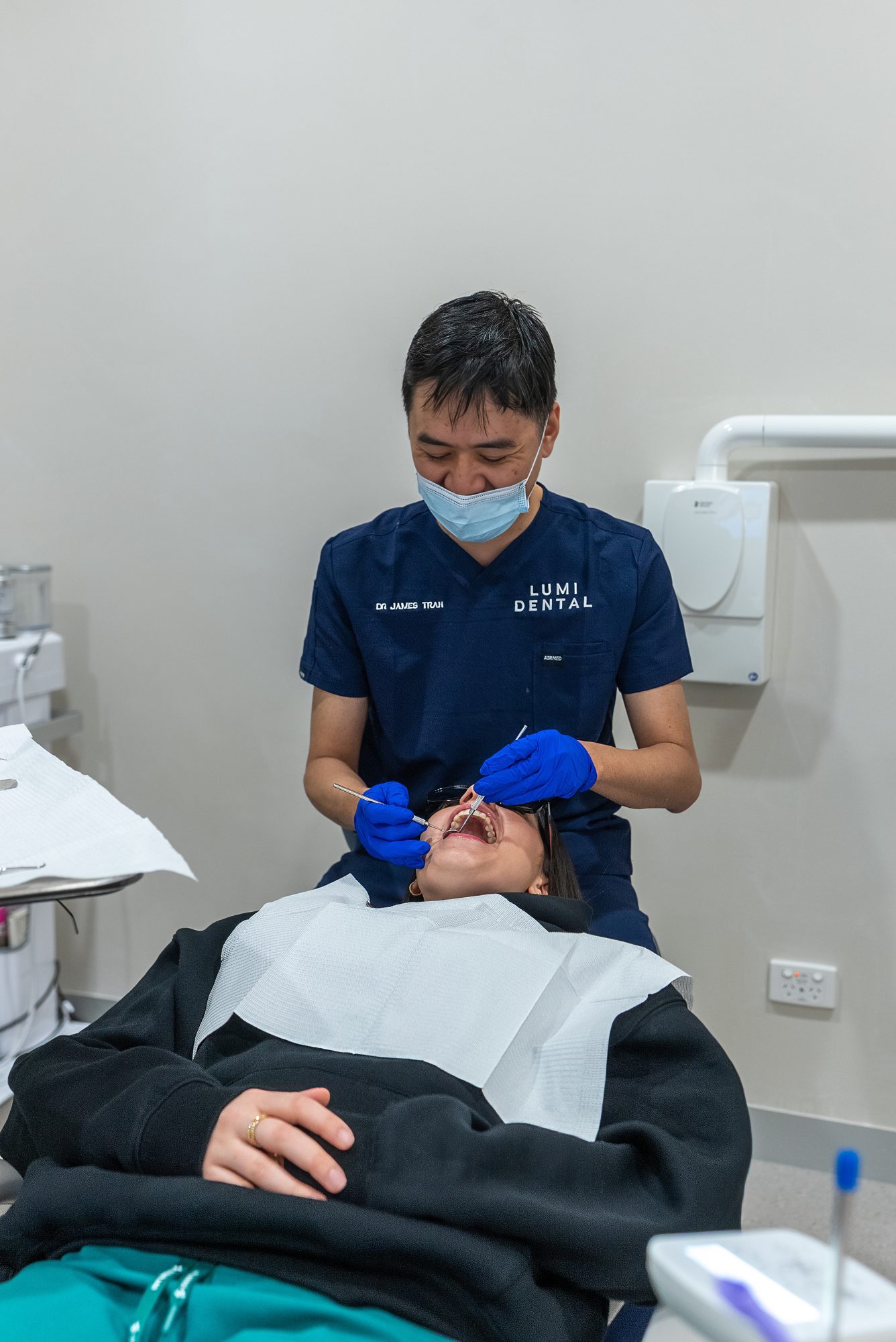
At Lumi Dental, a change in the gum around an implant starts with an examination by a dentist. Probing depths and bleeding points are recorded, the gum thickness and type are noted, and a new X-ray is compared with the baseline, using our in-house 3D X-ray where the shape of the bone matters. Our oral health therapists carry out cleaning and gum therapy around implants. Soft tissue grafting and advanced bone loss may be referred to a registered specialist.
Common questions
Is recession around an implant a sign it is failing?
Usually not on its own. Recession with stable bone and no bleeding is a gum problem around a sound implant. Recession together with bleeding, pus and bone loss is peri-implantitis, which can lead to loss of the implant if it is left untreated, so the X-ray comparison is what settles it.
Why does my implant look grey at the gum?
Most often because the gum is thin and the abutment beneath the crown shows through. If the gum is not bleeding or sore and the X-ray has not changed, it is a cosmetic matter.
Can the exposed collar be covered?
Sometimes. A soft tissue graft can cover it when the bone underneath is in good shape. Otherwise a new crown can be designed to hide it.
Concerned about the gum around your implant?
If the gum around an implant has changed colour, moved or started to bleed, we can probe the area, compare a new X-ray with your earlier one and explain which situation applies. Contact Lumi Dental in Melrose Park to book a free implant consultation (see current offers), or read more about dental implants at the practice.
Sources
- Galarraga-Vinueza ME and colleagues. Prevalence, incidence, systemic, behavioral, and patient-related risk factors and indicators for peri-implant diseases: an AO/AAP systematic review and meta-analysis. Journal of Periodontology, 2025
- Tavelli L, Barootchi S. Prevalence, incidence, risk, and protective factors for soft tissue dehiscences at implant sites in the absence of disease: an AO/AAP systematic review. Journal of Periodontology, 2025
- Berglundh T and colleagues. Peri-implant diseases and conditions: consensus report of workgroup 4 of the 2017 World Workshop. Journal of Periodontology, 2018
- Cai H and colleagues. Quantitative discoloration assessment of peri-implant soft tissue around zirconia and other abutments with different colours: a systematic review and meta-analysis. Journal of Dentistry, 2018
- Australian Dental Association (teeth.org.au): Gum disease treatment
About the author
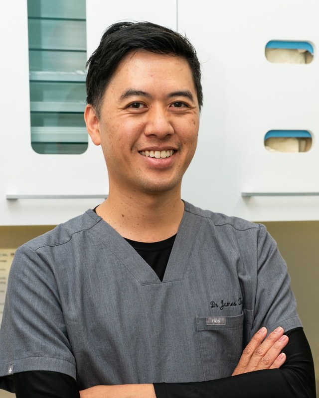
Dr James Tran is a general dentist at Lumi Dental in Melrose Park. He holds a BDent from the University of Sydney and a Graduate Diploma in Oral Implants, and is a Master of the International Congress of Oral Implantologists (MICOI).
This article is general information only and is not a substitute for an examination and individual advice from a registered dental practitioner. Suitability for any treatment varies from person to person.