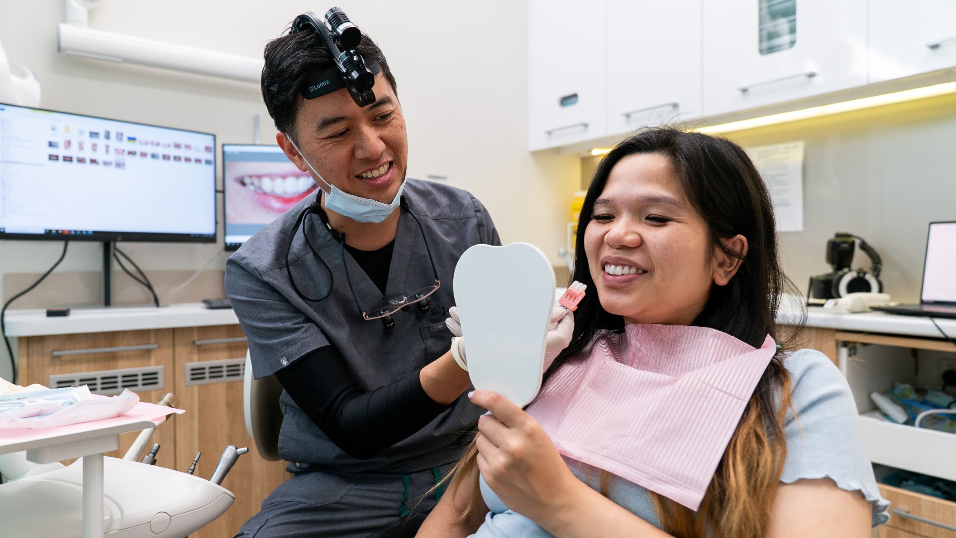
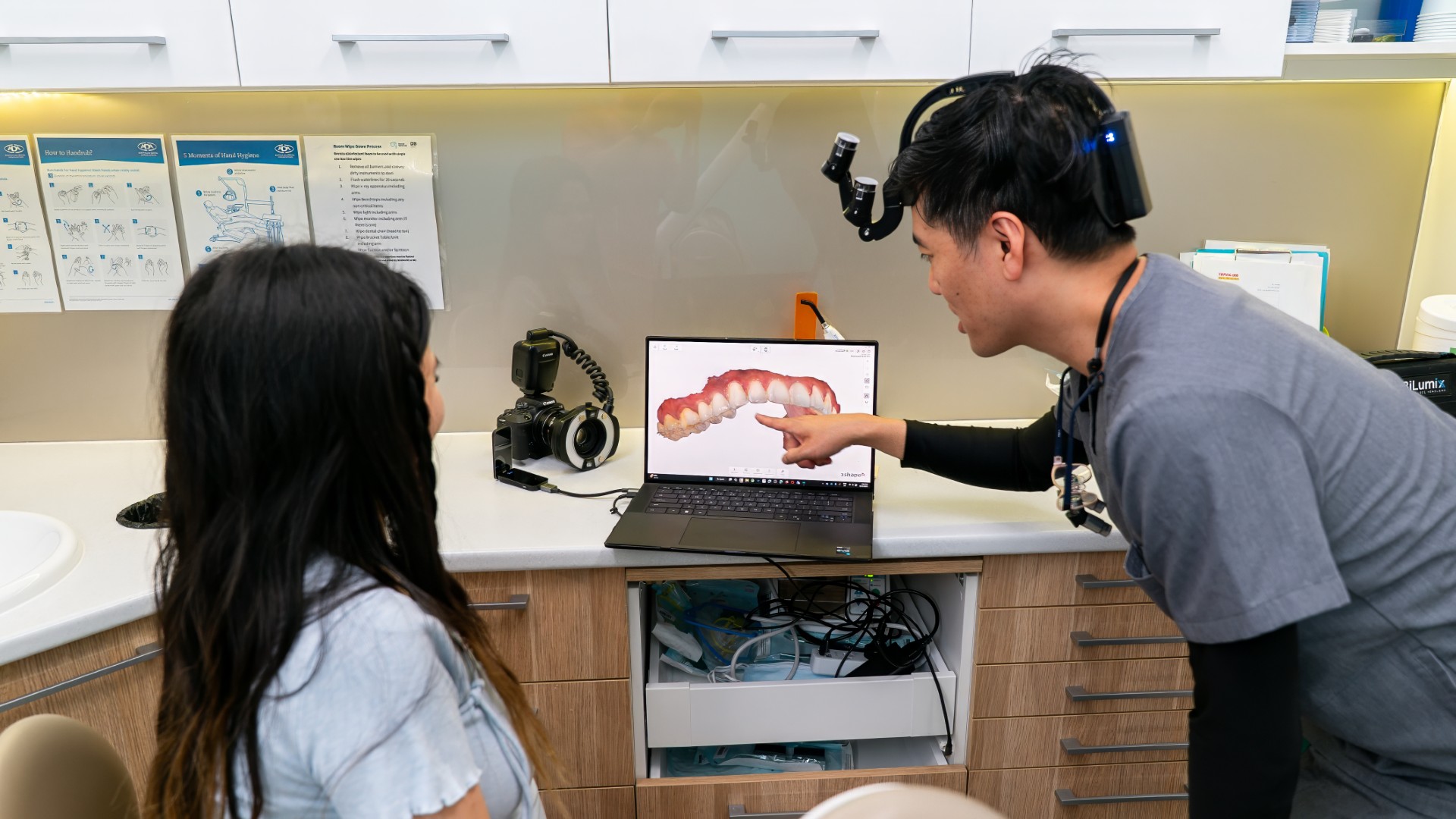
If you are wondering why teeth look different in photos, the usual answer is the camera rather than your teeth. A flash produces a hard mirror-like reflection off the enamel surface that you never see in a bathroom mirror, and automatic white balance quietly shifts the colour of everything in the frame. There is one exception worth taking seriously, and it is the reason this page exists: a photo is a poor way to judge your overall shade, but a good way to spot a single tooth that has changed.
Key takeaways
- Most of a tooth's colour comes from the dentine underneath, seen through semi-translucent enamel.
- Flash maximises surface reflection, which is why teeth can look unnaturally bright and flat in one photo and yellow in the next.
- Automatic white balance on a phone compensates for yellow with blue, so it actively alters the thing you are trying to assess.
- Two colours can match under one light and clash under another, which is why a result can look right in the surgery and wrong at home.
- Lip colour changes perceived tooth whiteness without changing the teeth at all.
- One tooth that is darker than the rest is a genuine finding and is worth having assessed.
What actually gives a tooth its colour
Enamel is not white in the way paint is white. It is semi-translucent, and most of what you perceive as tooth colour is the yellowish-grey dentine underneath, seen through it. Thinner or more translucent enamel lets more of that dentine colour through, so the tooth reads darker or yellower. At the biting edges of the front teeth there is often no dentine behind the enamel at all, so those edges photograph grey against the dark background of the mouth. That is normal anatomy, not a stain, and we cover it further in translucent teeth and what causes them.
Four things happen when light hits a tooth. Some passes straight through, some bounces off the surface as a mirror-like specular reflection, some scatters off the surface diffusely, and some is absorbed and scattered inside the tooth tissues. That second one, the specular reflection, is what a flash exaggerates. It is a reflection of the light source, not a property of your teeth, and it can wash out the surface completely.
Why dentists use cross-polarised photography
There is a whole technique built around removing that reflection. Cross-polarising filters cancel specular reflections from the tooth surface, saliva and restorations, reduce colour mismatch between light sources, and correct the way a direct flash overstates white patches on enamel. It is the recommended approach for photographing a shade to send to a laboratory.
Worth being precise about what it does. It removes a reflection so the underlying colour is visible. It does not measure your tooth colour, and nothing does, because there is no gold standard for the true colour of a natural tooth. Natural teeth vary more than any shade guide contains, which is why matching in the mouth is harder than in a laboratory.
What your phone is doing without telling you
Automatic white balance is the main problem. Clinical work on smartphone shade selection explicitly recommends turning it off, because the camera compensates: yellow tooth shades get pushed towards blue, and red gum tissue gets pushed towards blue-green. The phone is trying to make the whole scene look neutral, which means it is adjusting the exact quality you are trying to judge.
On top of that sits computational photography. Modern handsets stack frames, remap tones, sharpen selectively and sometimes apply skin and smile processing before you see the image. That processing is proprietary and changes with software updates, so nobody can tell you how much a given phone shifts tooth appearance, and anyone claiming a phone adds a set number of shades is making it up. Handset comparisons date almost immediately. What holds up is that phones still trail dedicated cameras on colour accuracy, even as newer models narrow the gap.
Lighting, colour temperature and metamerism
Shade assessment has a standard, and it is stricter than most rooms manage. The recommended condition is a light source between 5500 and 6500 kelvin with a colour rendering index above 90, where 5500 kelvin is roughly midday daylight under bright cloud. Studies have found most dental operatories sit below that temperature with a rendering index under 90.
Metamerism is the concept that explains the complaint people actually have. Two things can match perfectly under one light source and visibly clash under another. It is why a new filling or crown can look invisible in the surgery and obvious under the warm downlights of a restaurant, and why a whitening result can look one way at the clinic and another in your bathroom. Mixing light sources, daylight through a window plus an overhead LED plus a phone flash, makes it worse.
Everything around your teeth changes how white they look
Perceived whiteness is relative. Your visual system judges a colour against what surrounds it, an effect called simultaneous contrast. Teeth read as whiter against cool-toned lip colours, and a bluish undertone counteracts yellow undertones in enamel. Lip colour genuinely changes how white teeth appear, with no change to the teeth themselves.
The same applies to a tan, dark clothing near the face, and the background of the photo. It is how vision works, and worth knowing before you conclude something changed between two photos taken a week apart.
Is it the photo, or is it your tooth?
| What you are seeing | Usually the camera or lighting | Usually a real change in the tooth | What to do |
|---|---|---|---|
| All teeth look yellower than you remember | Warm indoor light, or white balance compensating | Gradual surface staining over years | Compare in daylight without flash before deciding |
| All teeth look unnaturally bright and flat | Direct flash and specular reflection | Rarely | Ignore the photo, it is showing the light source |
| Edges of the front teeth look grey or see-through | Dark oral background behind translucent enamel | Enamel wear or erosion, if it is progressing | Mention it at your next check-up |
| One tooth is darker than the rest | Almost never | Yes, this is the one that matters | Book an assessment |
| A tooth turned grey after a knock | No | Possible pulp damage, often painless | Have it examined, even without pain |
| Chalky white patches near the gumline | Flash exaggerates them | Often early demineralisation or a developmental defect | Assessment, since early lesions have options |
| Teeth look whiter with certain lip colours | Simultaneous contrast | No | Nothing, the effect is real but the teeth are unchanged |
| Whitening looked great at the clinic, dull at home | Metamerism and different light sources | Some rebound is expected | Reassess in daylight a couple of weeks later |
When it is not the photo
One tooth darker than the rest
This is where a photograph is genuinely useful. Lighting affects all your teeth at once. It does not single one out. So if one tooth is consistently darker or greyer than its neighbours across several photos taken in different places, that is a real difference and not a camera artefact.
Grey or dark-grey discolouration of a single tooth is the most common colour change after a knock, and the classic sign that the nerve inside has died. Blood vessels in the pulp rupture, blood pigment moves into the dentine tubules, and breakdown products of haemoglobin bind to the dentine. What catches people out is that this frequently causes no pain, so nothing prompts a visit.
Timing matters. A tooth that discolours within a few days of an injury has not necessarily lost its nerve, and some settle. Dark discolouration persisting for months is more likely to mean a dead nerve, and it can stay painless. That does not automatically mean root canal treatment, but it does mean the tooth needs examining and usually an x-ray. Our article on the types and causes of tooth discolouration covers the wider range of causes.
White spots are a separate question
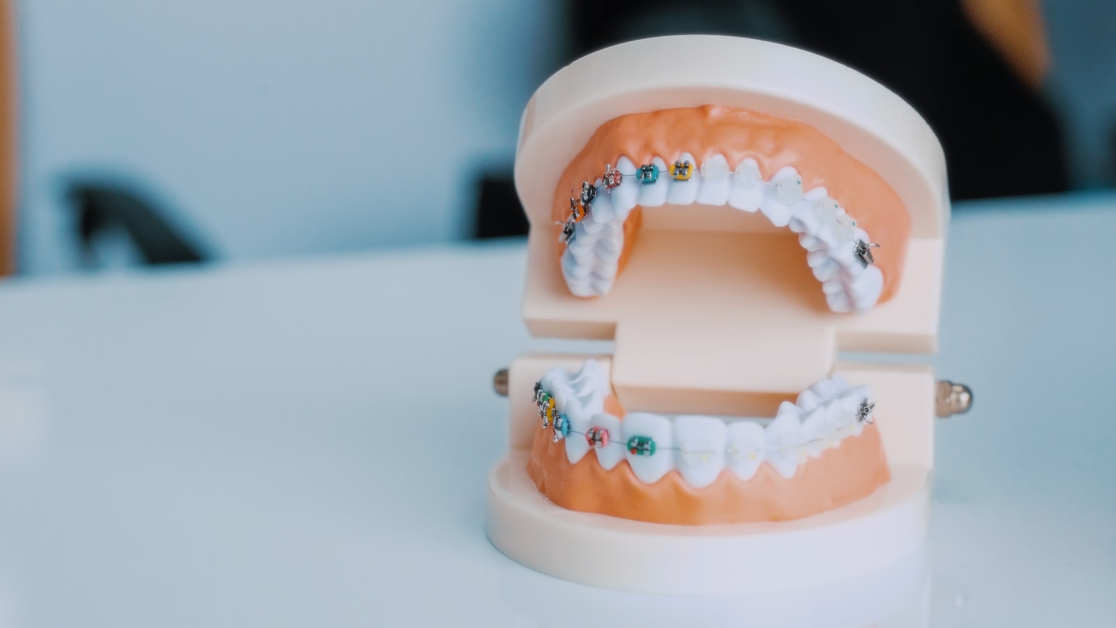
Chalky white patches show up strongly under flash, so people often notice them in photos long after they appeared. They are not a shade problem. The most documented cause is demineralisation around orthodontic brackets, where plaque sat for months, though developmental defects and fluorosis also produce them. Early demineralisation is a stage of decay, so it is worth assessing rather than covering up. We cover the options in white spots on teeth.
How a dentist takes a shade, and why not from your photo
A shade is recorded early in a visit, before the teeth dry out under a light and temporarily lighten. It is taken under controlled lighting, against a shade guide, and often photographed with polarising filters for the laboratory. Even then, agreement is imperfect. Repeatability between examiners has been measured at about 94 per cent for one modern bleached shade guide against about 55 per cent for an older classical guide, meaning that with the older system two clinicians disagree roughly half the time.
Instruments help but do not settle it. Spectrophotometers show clearly better repeatability and agreement between observers, while accuracy comparisons against the human eye go both ways depending on the study. Current practice combines an instrument reading with a controlled visual check. If you want the clinical side of this, our guide to tooth shade matching covers how a crown shade is chosen in the surgery, whereas this article is about what your own photos can and cannot tell you.
If you decide to whiten anyway
Measured properly, changes are gradual: one clinical series recorded a mean change of just over four shade tabs at two weeks and just under six at four weeks, with meaningful relapse after four weeks or more. In-office and dentist-supervised take-home bleaching produced approximately the same initial improvement, so the choice is more about convenience and sensitivity than results. How long it holds depends on habits, which we cover in how long teeth whitening lasts, and you can read about the process itself on our teeth whitening page.
One practical point: judge the result in daylight, not under a bathroom light and not in a photo with flash. And give it a fortnight, because teeth dehydrate during treatment and look lighter than they will settle at.
Common questions
Why do my teeth look yellow in photos but white in the mirror?
Bathroom mirrors are usually lit from above with warm light and you see your teeth at an angle, while a camera adds a flash, a different white balance and automatic colour correction. The phone often compensates for yellow tones by shifting the image, which can exaggerate rather than reduce them. Daylight without flash is the fairer comparison.
Does camera flash make your teeth look whiter?
It can, by producing a strong mirror-like reflection off the enamel surface that hides the underlying colour and flattens texture. It can also do the opposite, exaggerating white patches and making translucent edges look darker. Flash tells you about the light source more than about your teeth, which is why dental photography uses polarising filters to remove that reflection.
Why does one of my teeth look darker than the others?
Lighting affects every tooth in the frame, so a single dark tooth is usually a real finding. The most common reasons are a nerve that has died after an old knock, a large old filling, or a tooth that has had root canal treatment. It often causes no pain at all, so it is worth having it examined rather than waiting for a symptom.
Why do my teeth look grey at the edges?
The biting edges of front teeth are often enamel alone, with no dentine behind them, so they are translucent and pick up the dark background of the mouth. That is normal anatomy for many people. It can become more noticeable if enamel is thinning through wear or erosion, so it is worth mentioning at a check-up if it is increasing.
What are the white spots on my front teeth?
Usually early demineralisation, a developmental enamel defect, or fluorosis. Spots that appeared after braces are typically demineralisation where plaque sat around the brackets. Because early demineralisation is a stage of decay rather than a cosmetic issue, it is worth having them assessed before deciding what to do about their appearance.
Getting a real answer instead of a photo
If a photo has been bothering you, the quickest way to settle it is a look under proper light. The team at Lumi Dental can tell you in a few minutes whether what you are seeing is the camera, general surface staining, or a single tooth that needs attention. New patients can book a check-up and clean under the current new patient special on our current deals page, or ask a question through the contact page.
This article is general information only and is not a substitute for individual dental advice. If a tooth has changed colour after an injury, please have it assessed even if it does not hurt.