A coronectomy is an operation where only the crown of a lower wisdom tooth is removed and the roots are deliberately left in the jaw. It is offered when the roots sit hard against the inferior alveolar nerve, the nerve that supplies feeling to the lower lip, chin and gum. Wisdom teeth are among the most commonly removed teeth in Australia, with tens of thousands of procedures each year, and lower third molars most often sit close to that nerve. A coronectomy is not a smaller version of the same operation. It is a deliberate trade.
Key takeaways
- A coronectomy removes the crown of a lower wisdom tooth and leaves the roots undisturbed next to the nerve.
- The trade is accepting a small chance of needing a second procedure later, in return for a much lower chance of permanent numbness.
- A systematic review reported nerve injury in about 0.59 per cent of coronectomies across 2,176 cases.
- Meta-analysis found coronectomy reduced the odds of nerve injury compared with full removal, with an odds ratio of around 0.14.
- Retained roots commonly migrate away from the nerve over the first year or two, which is usually a good thing.
- It is generally not suitable if the tooth is already infected, decayed into the root, mobile, or if you are immunocompromised.
How you know your roots are near the nerve
The inferior alveolar nerve runs through a bony canal along the length of the lower jaw. On a standard panoramic x-ray, certain signs suggest the roots and the canal are intimate rather than merely overlapping. Darkening of the root where the canal crosses it, interruption of the white lines outlining the canal, diversion of the canal, and narrowing of the root are all warning signs.
A panoramic x-ray is a flat image of a three dimensional problem, so those signs tell you something might be there without telling you where. A cone beam scan shows whether the canal sits on the cheek side, the tongue side, or directly under the roots, and whether the bone between them is intact. In my experience that distinction changes the plan more often than people expect, because a fair number of teeth that look terrifying on a flat film turn out to have a comfortable margin of bone.
Close contact on a panoramic x-ray is common, but true nerve injury after removal remains uncommon. The scan exists to sort one group from the other rather than to frighten you.
What the numbness actually feels like
People imagine numbness as an absence of feeling, and that is the mildest version. Altered sensation after nerve injury covers a wider range. It can feel like a dental anaesthetic that never wore off, or like pins and needles, or a burning that comes and goes, and in some cases an area that is unpleasant to touch.
The practical effects are what make it matter. Dribbling when drinking. Biting the lower lip without noticing. Difficulty shaving or applying makeup on one side. Food sitting in the lip and not being felt. There is also an emotional weight to it that is hard to convey, because it is on the face and it is constant.
Most altered sensation after wisdom tooth surgery is temporary and resolves within weeks to months. A minority persists beyond six months, at which point recovery becomes less likely. Reported temporary rates after conventional removal of high risk lower wisdom teeth run up to about 16 per cent, with permanent rates up to about 3.6 per cent. After a successful coronectomy, temporary altered sensation is reported in roughly 0 to 2.2 per cent, and permanent injury is rarely reported.
Coronectomy compared with full removal
Neither option is automatically correct. The decision depends on how close the roots are, why the tooth needs treating, your age and your medical history.
| Consideration | Coronectomy | Full removal |
|---|---|---|
| What is removed | The crown only, cut a few millimetres below bone level, roots left in place | The entire tooth, crown and roots |
| Main reason to choose it | Roots are intimate with the nerve canal on a CBCT scan | Definitive removal, nothing left to monitor |
| Risk of permanent altered sensation | Rarely reported, with nerve injury of about 0.59 per cent across a large systematic review | Reported up to about 3.6 per cent permanent in high risk cases, and up to about 16 per cent temporary |
| Chance of needing a second procedure | A minority need the retained roots removed later. Reported success ranges from about 62 to 100 per cent, with one series of 167 cases reporting about 93 per cent | Not applicable once the tooth is out |
| Healing time | Commonly a week of swelling and soreness, sometimes less bone removal than a deep full extraction | Similar, though deep roots can mean more bone removal and a longer recovery |
| Suitability if the tooth is infected | Generally unsuitable if actively infected, decayed into the root, or mobile | Usually the appropriate option in those situations |
| Follow-up imaging | X-rays over the first one to two years to watch root migration | Usually none beyond healing checks |
| Typical cost direction in the Australian market | Broadly similar to a complex surgical removal, often slightly higher, plus a CBCT scan commonly costing in the low hundreds of dollars | Surgical removal of an impacted lower wisdom tooth commonly ranges from a few hundred to well over a thousand dollars per tooth |
Costs vary widely between practitioners, and the anaesthetic choice matters as much as the surgery. Treatment under sedation costs more than treatment under local anaesthetic. If anxiety is part of your decision, our overview of nitrous oxide for adults explains the lighter end of the options, and intravenous sedation covers the deeper end.
The procedure, step by step
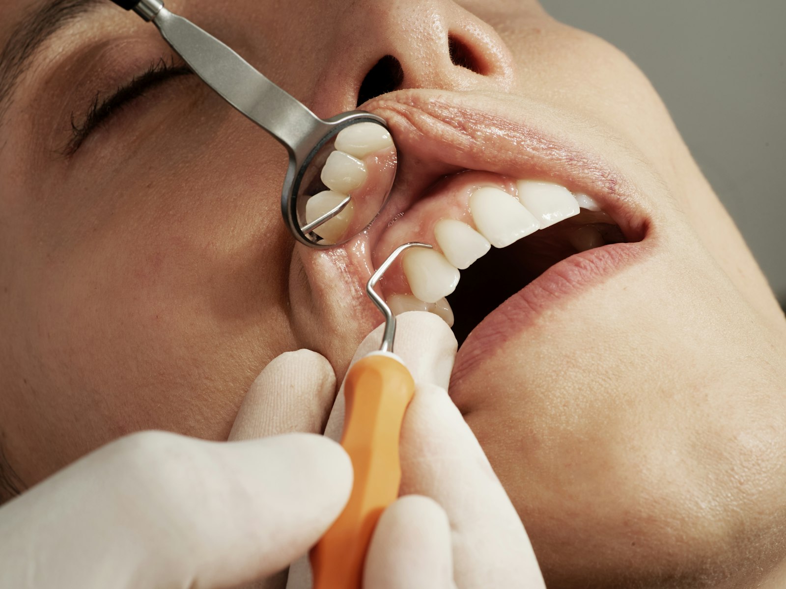
A coronectomy is usually done under local anaesthetic, with sedation available if you prefer. Most appointments run between 45 and 90 minutes.
- The gum is numbed thoroughly and a flap is lifted to expose the tooth.
- A small amount of bone over the crown is removed if needed.
- The crown is sectioned off, usually cutting through the tooth at an angle, and lifted away.
- The remaining root surface is reduced so it sits at least 3 mm below the bone crest, which allows bone to grow over it.
- The roots are left completely undisturbed. Critically, they are not loosened, because a mobilised root has to come out and the whole point of the operation is lost.
- The socket is irrigated, the flap is repositioned and sutures are placed.
Antibiotics are not routinely required for every case, and the decision follows the same principles used for any surgical extraction. If the tooth has been repeatedly inflamed around the gum flap, that history matters, and our guide to pericoronitis explains why recurrent episodes often push the decision towards treatment sooner rather than later.
Recovery and what to expect afterwards
Recovery is broadly similar to a surgical wisdom tooth extraction. Expect swelling that peaks around day two or three, jaw stiffness for several days, and soreness that eases through the first week. Sutures are usually reviewed at about a week.
The aftercare rules are the same ones that protect any surgical socket. No rinsing for the first 24 hours, no smoking, no straws, soft food, and gentle salt water rinses from day two. Our extraction recovery timeline sets out the normal day by day pattern, and the guide to dry socket covers the main early complication. Some patients also notice temporary tingling in the lip afterwards, usually from the surgery itself rather than a lasting injury, and it typically settles over days to weeks.
How root migration is monitored
The retained roots do not sit still. In the majority of cases they migrate upwards, away from the nerve canal, most rapidly in the first six to twelve months and then slowing. This is generally welcome, because a root that has moved away from the nerve can be removed conventionally if it ever needs to be.
Monitoring is usually a review x-ray at around six to twelve months, then again at about two years, and after that only if symptoms appear. Bone commonly grows over the root surface, and the roots often become buried and quiet.
Reintervention is needed in a minority of cases. The usual reasons are infection around the roots, roots that erupt through the gum, or persistent pain. Because the roots have usually moved away from the nerve by then, the second procedure is generally more straightforward than the original operation.
When a coronectomy is not the right choice
The operation depends on leaving healthy, sterile roots in a healthy jaw. Where that condition is not met, it should not be attempted.
- The tooth is actively infected, or there is an abscess on the roots.
- Decay extends into the root, so what is left behind would not be sound.
- The tooth is mobile, since loose roots cannot be left in place.
- The tooth is impacted in a way that makes safe sectioning impossible.
- The patient is immunocompromised, on chemotherapy, or has had radiotherapy to the jaws.
If you are on bone medicines such as alendronate or denosumab, that history changes the surgical planning, and our article on bone medicines and jaw healing explains why.
Frequently asked questions
Is a coronectomy safer than taking the whole wisdom tooth out?
For teeth with roots genuinely intimate with the nerve, published data suggest a substantially lower risk of nerve injury, with meta-analysis reporting an odds ratio of around 0.14 compared with full removal. That comes at the cost of retained roots and a possible second procedure. For teeth away from the nerve, full removal remains the straightforward option.
Will the roots left behind become infected?
Usually not. In most reported series the roots heal quietly with bone growing over them. Infection around retained roots is the main reason for later removal, and it occurs in a minority of cases. Regular check-ups and review x-rays over the first two years are how this is picked up early rather than late.
How long do the roots take to move away from the nerve?
Migration is fastest in the first six to twelve months and generally slows after that, continuing more gradually into the second year. Most movement is upwards, away from the nerve canal. This is why review imaging is usually scheduled at around six to twelve months and again near the two year mark.
Do I need a CBCT scan before a coronectomy?
A cone beam scan is commonly recommended when a panoramic x-ray shows signs of close contact between the roots and the nerve canal. It shows the true three dimensional relationship, including which side of the roots the canal sits on. That information is what determines whether coronectomy is the sensible option or whether full removal is safe.
Is a coronectomy more painful than a normal extraction?
Most people report a similar recovery, with swelling peaking around day two or three and settling over a week. Sometimes less bone needs removing than for a deep full extraction, which can make recovery slightly easier. Pain relief is managed the same way, usually with paracetamol and an anti-inflammatory.
Related reading: oral surgeon versus general dentist for extractions and implants.
The bottom line
A coronectomy exists for one specific situation, a lower wisdom tooth whose roots are wrapped around the nerve, where taking the whole tooth carries a real chance of permanent numbness. Leaving the roots is a considered exchange of a small chance of a second, easier procedure for a much lower chance of a permanent change to your lip and chin. Whether it applies to you depends on what the scan shows. Book an assessment and a written quote with the team at Lumi Dental, read more about general dental care in Melrose Park, and check the current offers before you decide. This article is general information and not a substitute for personal dental or medical advice.