Roughly a quarter of permanent teeth that are knocked go on to fill in part of their pulp space with extra dentine, and a tooth doing that often turns slowly yellower than its neighbours. That is the most common reason for one yellow front tooth that does not hurt. It can often be lightened on its own with a specially cut tray, but a vitality test and an X-ray come first, because colour alone cannot show whether the nerve inside is healthy.
Written by Dr James Tran, BDent (University of Sydney), MICOI, GradDip (Oral Implants), general dentist at Lumi Dental in Melrose Park.
A knock you may have forgotten
Enamel is fairly translucent, so much of a tooth's colour comes from the dentine underneath. The Australian Dental Association notes that an injury can lead a tooth to make more dentine to protect the pulp, and that injured teeth can darken over time. When this carries on for years, the pulp chamber and canal narrow, sometimes until they almost disappear on an X-ray. Dentists call this pulp canal obliteration.
It is common. A 2024 systematic review in Brazilian Oral Research (Abreu and colleagues), drawing on 34 studies, estimated that about 27.6% of injured permanent teeth develop it. A review in the International Endodontic Journal (McCabe and Dummer, 2012) describes the typical picture: the pulp space fades on the X-ray and the crown takes on a yellow tint.
The usual story is a bump to the mouth years earlier, perhaps in childhood sport, no pain at the time, and one tooth that has gradually drifted yellower. Other explanations include wear that has exposed dentine, an old filling that has darkened, or neighbouring teeth that were whitened years ago while this one kept its original shade.
The colour is the first clue
An even yellow suggests a living tooth with extra dentine. Grey, blue-grey or dark brown more often points to a tooth whose nerve has died, with blood pigments left in the dentine, or to an old root canal filling. Brown or white patches that have been there since the tooth came through formed while the enamel was developing, and those behave differently again.
| What you see | Common explanation | Typical next step |
|---|---|---|
| Even yellow, no pain | Extra dentine after an old knock; wear; a darkened filling | Vitality test and X-ray, then possibly single-tooth whitening |
| Grey, blue-grey or dark brown | Nerve has died, or an old root canal filling | Root canal treatment if needed, then whitening from inside the tooth |
| Brown or white patch since the tooth came through | Enamel that formed abnormally (hypoplasia or fluorosis) | Options such as resin infiltration, microabrasion, bonding or a veneer |
| Darker only near the gum | Surface stain, exposed root or a filling showing through | Professional clean, then reassess |
| Both canines darker than the front teeth | Usually normal; canines tend to be a little darker | Whole-mouth whitening if wanted |
The grey route is covered in our guides to grey teeth, signs of a dead or dying tooth and internal bleaching. The ADA explains that stains inside a tooth are very difficult to shift from the outside, which is why a root-treated tooth is usually whitened from within. Developmental patches are explained in our article on enamel hypoplasia; whitening gel often brightens the tooth around a patch first, so the patch can look more obvious for a while.
Why the X-ray comes before any gel
A tooth with a narrowed pulp space usually stays healthy. McCabe and Dummer report that only about 7% to 27% of teeth with pulp canal obliteration go on to develop a dead nerve with signs of infection at the root tip. But a tooth with a dying nerve, a crack or decay will not respond well to whitening, may become sore with peroxide, and needs treatment that gel would only postpone.
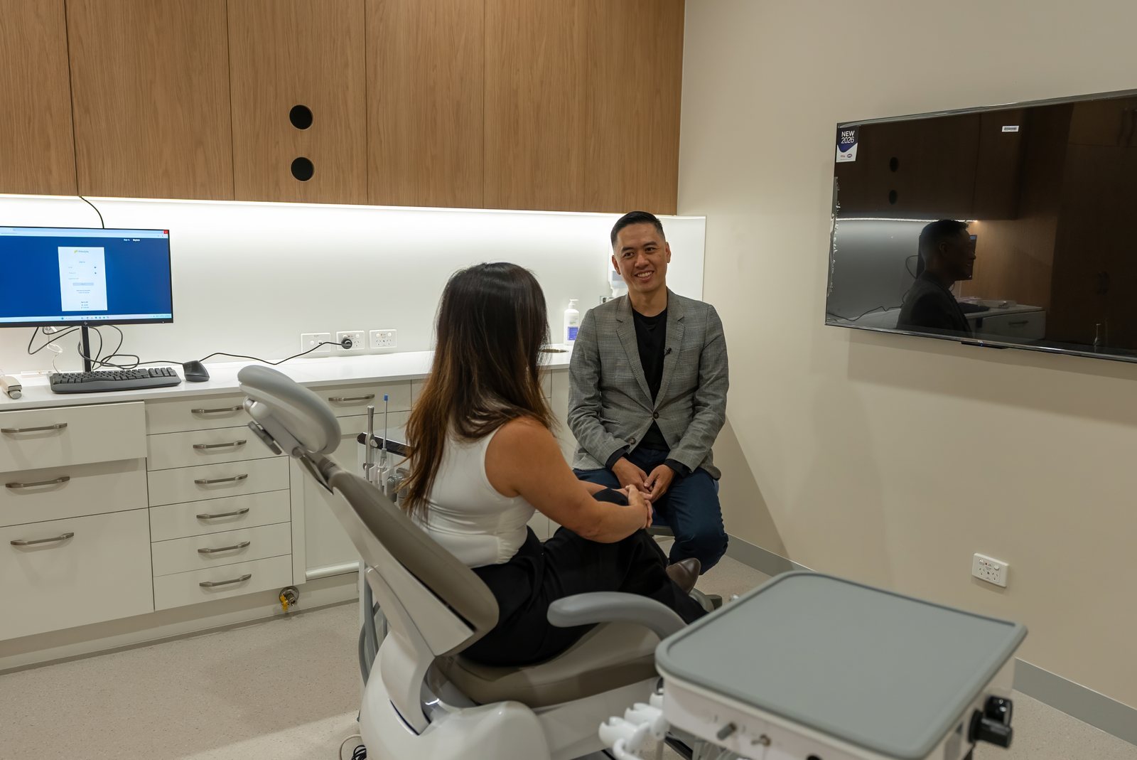
Standard cold tests are also unreliable in these teeth, because the extra dentine dulls the response. So the dentist uses the test alongside a small X-ray, which shows the narrowed canal, checks for a shadow at the root tip and looks for fractures. A photograph with a shade guide records the starting colour, and the bite is checked for wear that might keep going if its cause is not managed.
How a single-tooth tray works
If the tooth is alive and sound, it can be whitened with a take-home tray that has been modified for one tooth. A protocol described in Dentistry (UK) in 2007 sets out the usual approach. A full tray is made first. Small windows are then cut on either side of the yellow tooth, so the gel stays against that tooth instead of spreading to its neighbours, which would lighten faster and keep the gap.
Treatment runs in two stages. The whole arch is whitened first for about two weeks with a milder carbamide peroxide gel. A stronger, dentist-supplied gel is then used on the single tooth for around four to six weeks, sometimes with a further two weeks. Progress is slow because the extra dentine is densely packed, although the protocol reports little or no sensitivity in these living teeth.
Reviews every two to three weeks let the dentist compare the tooth with the shade guide and decide when to stop, because a single tooth can overshoot and end up lighter than the rest. Not every tooth reaches a full match, and no particular shade can be promised. The ADA lists sensitivity to cold and irritation or chemical burns where gel touches the gums as the main side effects, which a well-fitting tray helps keep down.

If the tooth does not catch up
Some teeth stop short of their neighbours, especially with very dense dentine, a grey undertone or a large old filling. The next step is to cover the surface: composite bonding for a modest difference or a worn edge, or a single porcelain veneer where the mismatch is larger. Our guide to a single veneer on one front tooth explains how the colour is matched.
Whiten first and match any restoration afterwards, usually after waiting about two weeks for the shade to settle. Whitening gel does not change the colour of porcelain crowns, veneers or white fillings, as the ADA points out. Our article on whitening with fillings or bonding covers the timing.
Checking and whitening one tooth at Lumi
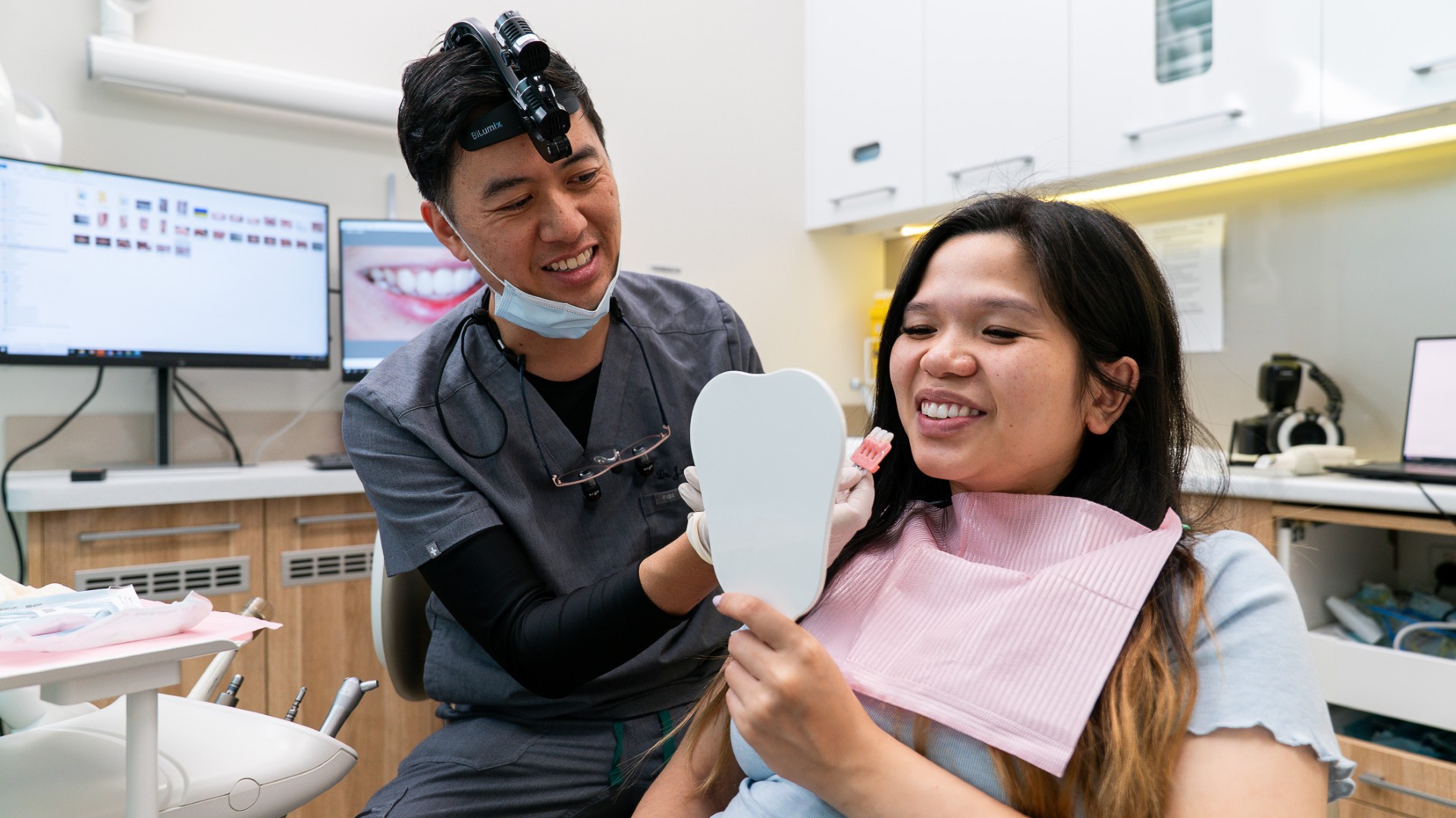
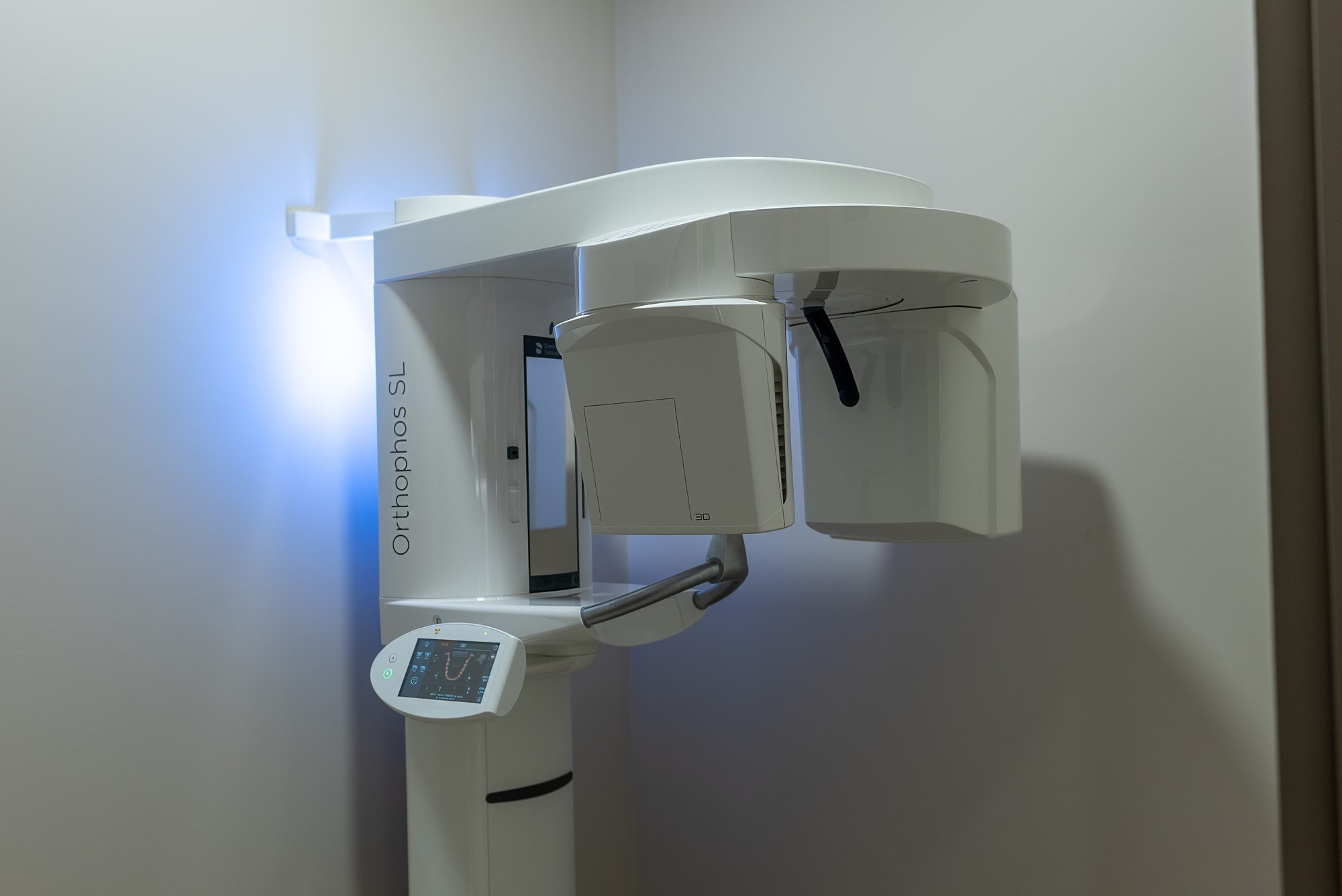
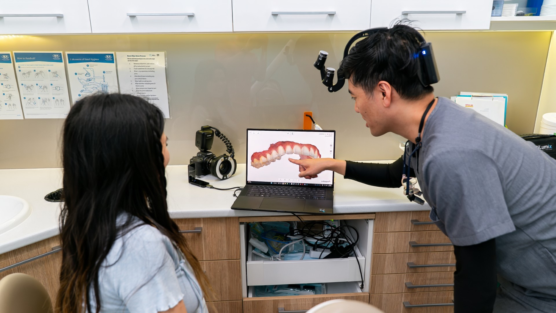
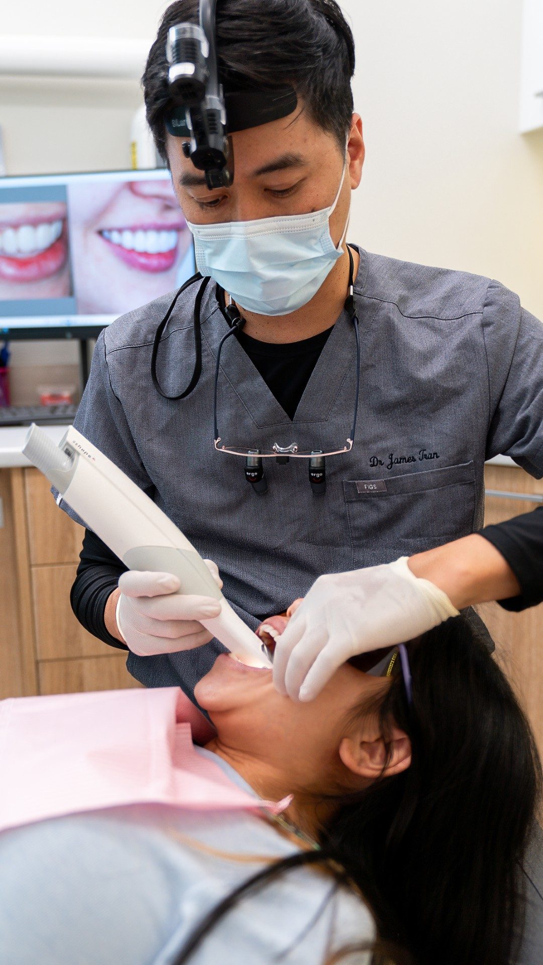
At Lumi Dental, a single discoloured tooth starts with an examination by a dentist, a vitality test and an X-ray taken on site. Clinical photographs record the starting shade, and our 3D intraoral scanner is used instead of impressions when a tray is made. Our oral health therapists can remove surface stain first so the tooth's real colour shows.
Lumi Home is our take-home whitening with custom trays and Boutique gels, offered as an overnight gel for sensitive teeth or a daytime gel for teeth that are not sensitive. Lumi Refresh and Lumi Radiance combine in-chair whitening with Boutique take-home whitening. We do not promise a particular shade. The teeth whitening page explains how a consult runs. Complex root canal cases may be referred to a registered specialist.
What patients ask about one yellow tooth
Can you whiten just one tooth?
Often, if the tooth is alive and sound. A take-home tray is cut so the gel reaches only that tooth, with reviews every few weeks. A tooth whose nerve has died is usually whitened from the inside instead.
How long does it take?
Longer than a normal course. The published protocol describes about four to six weeks on the single tooth after a two-week whole-arch stage, and sometimes longer.
Why are my canines more yellow than my front teeth?
Canines are naturally a little darker than the front teeth in most people. If both match each other, whitening the whole arch lifts them with the rest. If only one is darker, it is a single-tooth question.
Will whitening fix a tooth that looks dark because of an old filling?
No. Gel lightens natural tooth, not composite, porcelain or amalgam. The usual approach is to whiten first and replace the filling afterwards.
One tooth standing out? Start with a free consult
If one tooth has drifted away from the others, the team at Lumi Dental in Melrose Park can test it, take an X-ray and explain your options. Book a free cosmetic consult or read about teeth whitening at Lumi Dental.
Sources
- Australian Dental Association (teeth.org.au): Teeth staining
- Abreu MGL and colleagues. Prevalence of pulp canal obliteration after traumatic dental injuries: a systematic review and meta-analysis. Brazilian Oral Research, 2024
- McCabe PS, Dummer PM. Pulp canal obliteration: an endodontic diagnosis and treatment challenge. International Endodontic Journal, 2012
- Dentistry.co.uk: Single vital tooth whitening (2007)
- Australian Dental Association (teeth.org.au): Teeth whitening
About the author

Dr James Tran is a general dentist at Lumi Dental in Melrose Park. He holds a BDent from the University of Sydney and a Graduate Diploma in Oral Implants, and is a Master of the International Congress of Oral Implantologists (MICOI).
This article is general information only and is not a substitute for an examination and individual advice from a registered dental practitioner. Suitability for any treatment varies from person to person.